Introduction
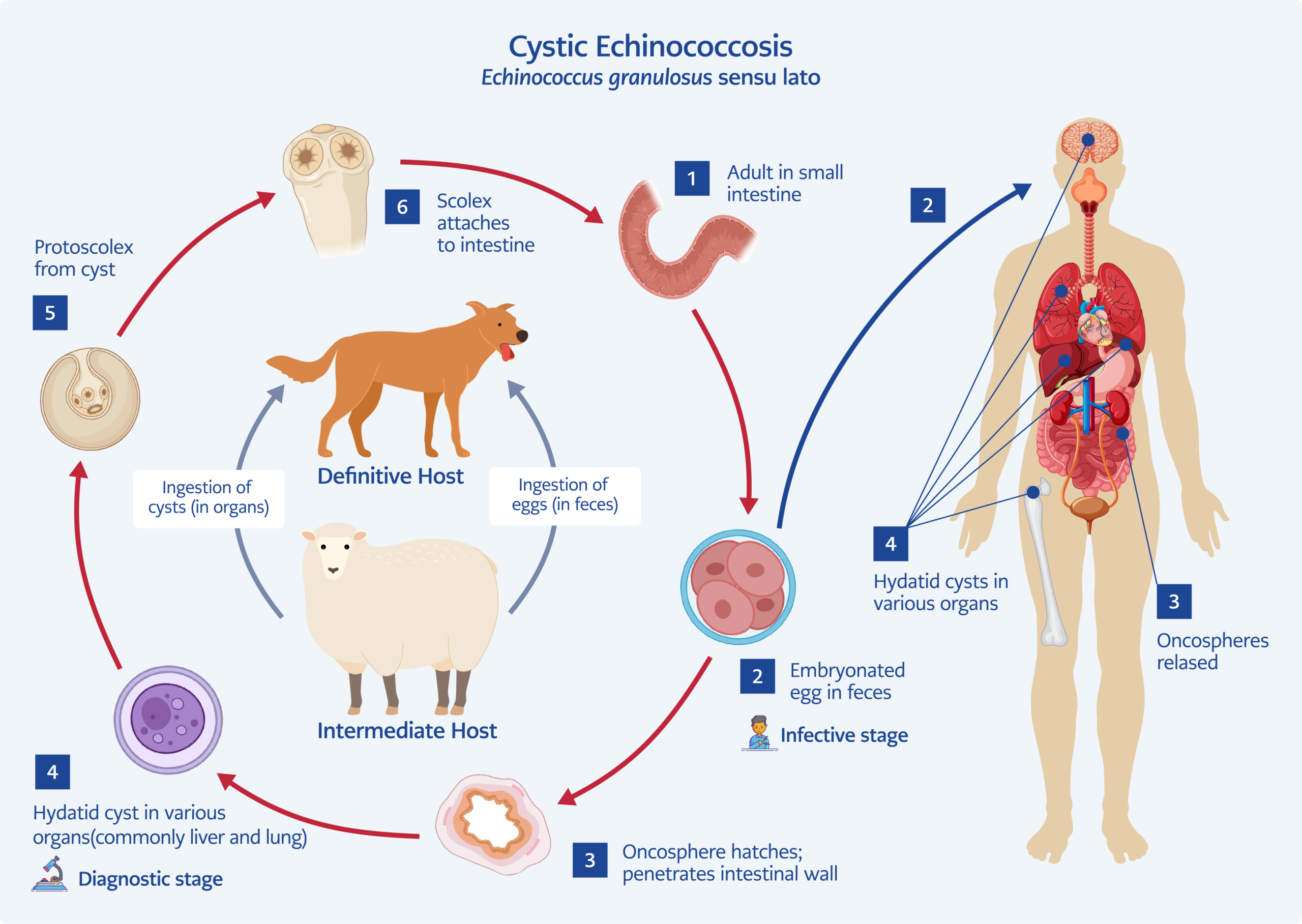
Figure 1. Life cycle of cystic echinococcosis. Adapted from reference 6.
Hydatid disease is a parasitic infection caused by Echinococcus granulosus and Echinococcus multilocularis (1). The life cycle of E. granulosus involves two main hosts: an intermediate host and a definitive host. Dogs are the most common definitive hosts; adult worms attach to the intestinal mucosa, and eggs are excreted in the feces. In intermediate hosts (humans and herbivores such as cattle, sheep, goats, camels, horses, and pigs), the eggs hatch and the larvae can reach various organs via the circulatory system (Figure 1). This disease predominantly affects the lungs and liver, while involvement of the musculoskeletal system occurs in approximately 4% of cases (2,3).
Immunosuppressed individuals, people living in rural areas, and those who have contact with domestic animals are at increased risk (4). In endemic regions such as South America, Eastern Europe, East Africa, Central Asia, China, and Russia, hydatid disease remains relatively common, with an annual incidence ranging from <1 to 200 cases per 100,000 population (1–5).
The diagnosis of hydatid disease is based on clinical evaluation combined with immunological, radiological, and histopathological findings. Due to the rarity of musculoskeletal involvement and occasional confusion with tumors, delays in diagnosis and treatment may occur. Surgery has remained the cornerstone of treatment for large cysts, superficial or potentially rupturable cysts, infected cysts, and cysts located in vital anatomical regions or causing significant mass effect (7,8). The management of musculoskeletal hydatid disease is particularly challenging for orthopedic surgeons because of the risks of recurrence and limb loss. There are limited data in the literature regarding the optimal management of such cases (7). Although preoperative albendazole therapy can reduce the need for aggressive surgical procedures, the recommended treatment for musculoskeletal hydatid cysts is wide excision to prevent cyst rupture and the dissemination of protoscoleces (3,7,8). In cases where complete cyst removal is not possible, alternative treatment options include cyst drainage, complete removal of the germinative membrane, and irrigation of the cyst cavity with a scolicidal solution (9).
In this study, we evaluated the treatment outcomes of patients with musculoskeletal hydatid cysts in our endemic region who could not be treated with wide excision because of cyst location and were treated with surgical drainage followed by the application of povidone-iodine as a scolicidal agent.
Materials and Methods
Study Design and Setting
Patients with musculoskeletal hydatid disease treated between 2016 and 2024 were included in the study. The final study cohort consisted of patients who met the inclusion criteria and underwent controlled cyst drainage and povidone-iodine sterilization. Written informed consent for publication was obtained from all patients. The study was conducted in accordance with the Declaration of Helsinki. Ethics approval was obtained from the University Ethics Committee (Approval No: 30.04.2026-190).
Inclusion and Exclusion Criteria
Patients with osseous involvement, those treated with chlorhexidine as the scolicidal agent, those who underwent wide excision, or those with inadequate follow-up were excluded from the study. Patients in whom wide excision was not feasible because of the risk of surgical morbidity were included and treated with controlled cyst opening, mechanical debridement, and chemical sterilization using povidone-iodine.
Wide excision was considered unfeasible when complete en bloc excision was expected to result in substantial functional impairment, major neurovascular injury, excessive soft-tissue sacrifice, or when anatomical constraints prevented safe resection with adequate margins. Surgical feasibility was assessed preoperatively based on magnetic resonance imaging (MRI) findings and multidisciplinary evaluation by orthopedic surgeons, radiologists, and infectious disease specialists. In the present series, this situation was most commonly encountered in lesions located in the hand and wrist, where wide excision could compromise tendon function, joint mobility, or digital function. Similarly, lesions involving the iliacus muscle and deep pelvic regions were considered unsuitable for wide excision because of their proximity to major neurovascular structures and the technical difficulty of achieving safe en bloc excision. In addition, if recurrence were to occur, repeat surgery in these anatomically complex regions would likely be associated with substantial morbidity.
Preoperative Evaluation and Surgical Technique
Before surgical treatment, patients were radiologically screened with ultrasonography and chest radiography to assess lung, liver, and other organ involvement. All patients were evaluated by the infectious diseases department, and serological testing, including serum immunoglobulin G (IgG) against E. granulosus, assessed by immunofluorescence antibody testing, was positive in all patients. In addition, eosinophilia was detected in peripheral blood samples in all cases. All patients received oral albendazole at a dose of 400 mg/day for two weeks preoperatively and continued the same regimen for three months postoperatively.
During surgery, the cyst was carefully opened, and its contents were completely evacuated by aspiration. The cyst cavity was then filled with 10% povidone-iodine solution and left in situ for 20 minutes to achieve chemical sterilization. Subsequently, the solution was reaspirated, and the cavity was carefully inspected for residual daughter cysts. After confirming complete evacuation, the cyst wall was excised. The video demonstrating the surgical technique is available from the corresponding author upon request.
Postoperative follow-up consisted of regular clinical evaluations and plain radiographs. Patients were evaluated monthly for the first three months, every three months until the end of the first postoperative year, and every six months thereafter. Magnetic resonance imaging was performed early if patients reported symptoms suggestive of recurrence, such as pain, swelling, or local pressure sensation in the surgical area. All patients underwent MRI at the end of the first year, regardless of symptoms. Recurrence was defined as the presence of a new cystic lesion at the surgical site detected on MRI, with or without accompanying clinical symptoms.
Statistical Analysis
Descriptive statistics were used to evaluate the patients’ demographic data and lesion locations. Continuous variables were presented as mean (range), while categorical variables were presented as frequencies (n) and percentages (%). Statistical analyses were performed using IBM SPSS Statistics for Windows, Version 20.0 (IBM Corp., Armonk, NY, USA).
Results
Of the 35 patients with musculoskeletal hydatid disease treated between 2016 and 2024, 28 were excluded from the study. Eleven patients underwent wide excision, eight patients had inadequate follow-up, six had osseous involvement, and three were treated with chlorhexidine as the scolicidal agent. The final study cohort consisted of seven patients.
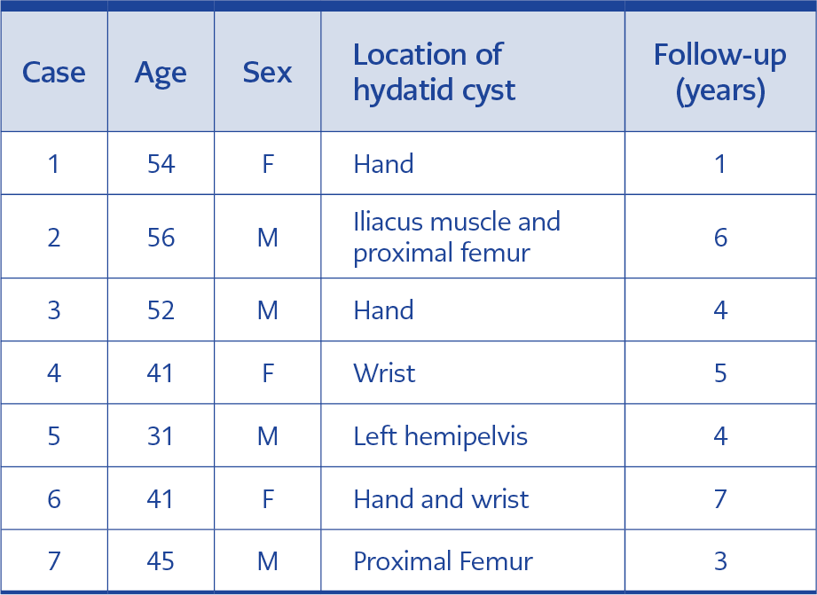
Table 1. Demographic and clinical characteristics of
patients with musculoskeletal hydatid disease.
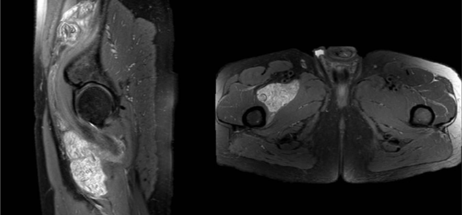
Figure 2. Magnetic resonance imaging of a patient with hydatid cysts involving the right iliacus muscle and proximal thigh.
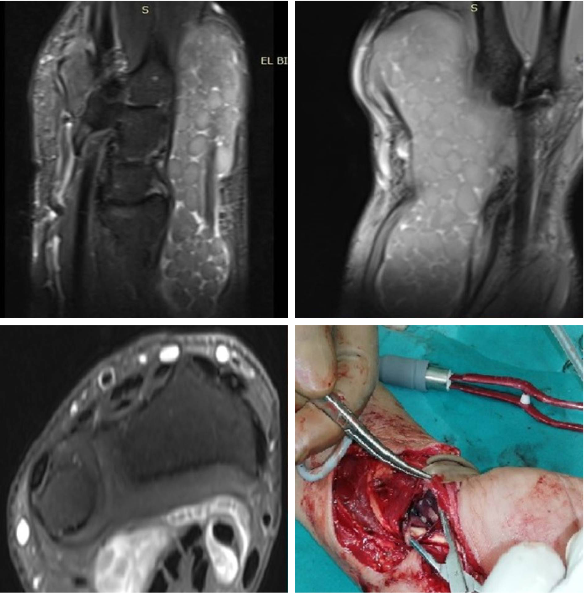
Figure 3. Magnetic resonance imaging and intraoperative images of a patient with a hydatid cyst of the wrist.
The mean age of the patients was 45.7 years (range, 31–56 years), with four males and three females. The mean follow-up period was 4.2 years (range, 1–6 years), and the most common presenting complaint was chronic pain partially interfering with daily activities (Table 1). Lesions were located in the right iliacus muscle, the left hemipelvis, the proximal right thigh (Figure 2), and the hand and wrist region in four patients (Figure 3).
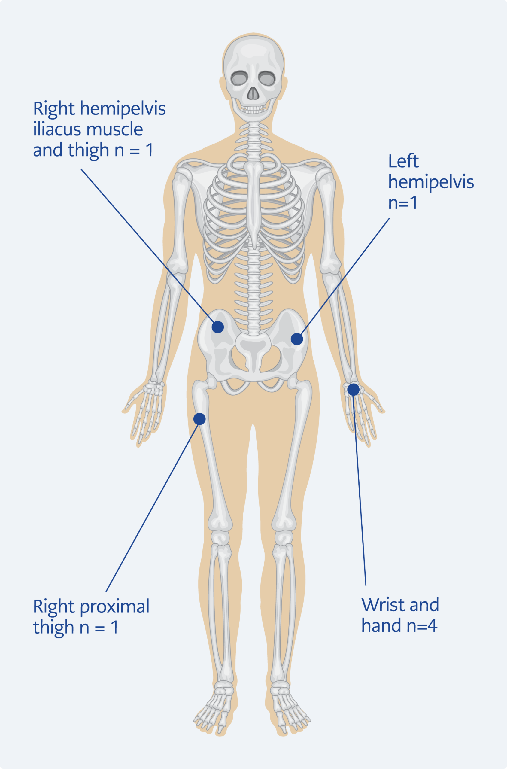
Figure 4. Anatomical distribution of musculoskeletal hydatid cysts in the study patients.
The locations of the patients’ lesions are illustrated in Figure 4.
All patients were evaluated preoperatively in collaboration with the radiology team. This surgical approach is used in patients for whom wide excision was deemed unfeasible because of the risk of limb function loss, neurovascular injury, or, for lesions involving the hemipelvis, because complete en bloc excision was not feasible. One patient had lesions in the lungs consistent with hydatid disease and underwent surgical treatment by the thoracic surgery team. No perioperative anaphylaxis was observed. Postoperatively, no patient developed a surgical site infection. At postoperative follow-up, serological testing remained positive in 80% of patients. Follow-up evaluations demonstrated complete resolution of pain and swelling.
At the final follow-up, all patients had returned to their preoperative daily activities without clinically significant functional limitations. In patients with hand and wrist involvement, no limitations in finger motion, wrist range of motion, or hand function interfering with daily activities were observed. No deficits were identified on muscle strength examination of the affected extremity in any patient. Furthermore, no patient required additional surgical intervention or physiotherapy for functional impairment.
Discussion
Hydatid cysts in the musculoskeletal system are rare, and therefore only a limited number of cases have been reported in the literature. This rarity may be due to the presence of lactic acid in the muscles, which may create an environment that inhibits the development of hydatid disease (10,11). Preoperative diagnosis of hydatid disease is challenging and relies on imaging findings and serological tests. However, serological tests, particularly for E. granulosus, have limited sensitivity or specificity. A high index of suspicion is especially required in patients living in or traveling to sheep-rearing regions where hydatid disease is endemic.
The majority of these patients present late because of delays in diagnosis, often with markedly enlarged cysts. Consequently, the recommended surgical approach for musculoskeletal hydatid disease—wide excision—may lead to functional loss in the affected extremity, particularly for lesions located in the hand and wrist, as observed in our patient series, and in some cases may not be feasible. Under certain circumstances, controlled cyst drainage followed by irrigation with scolicidal agents may represent an alternative treatment option (12,13).
In cases where complete excision of a soft tissue–localized cyst is not possible, various scolicidal agents—including formalin, hypertonic saline, cetrimide, chlorhexidine, hydrogen peroxide, povidone-iodine, and ethanol—have been used, but many have been abandoned because of adverse effects. Povidone-iodine (10%) and chlorhexidine gluconate are effective scolicidal agents that have been used in hydatid cyst surgery for many years. In musculoskeletal hydatid disease, the use of scolicidal agents has been limited and has primarily been described in case reports (14). Chlorhexidine and povidone-iodine are the most commonly used scolicidal agents, but evidence regarding their use remains limited. Most reports involve patients with liver or abdominal involvement, in whom these agents have been used by general surgeons with favorable outcomes (11,15,16).
Cyst opening can lead to anaphylaxis and the dissemination of daughter cysts, resulting in the formation of new lesions. When this technique is used, recurrence rates of up to 80% have been reported for osseous hydatid cysts (17). To reduce this risk and prevent systemic dissemination, preoperative and postoperative antiparasitic drug therapy is recommended. Benzimidazole derivatives (albendazole and mebendazole) are the most commonly used agents (18). Albendazole is preferred because of its better absorption. The optimal treatment regimen remains a matter of debate. Typically, the drug is administered in 28-day cycles at a dose of 15 mg/kg/day, followed by a 14-day drug-free interval. One cycle is given preoperatively, and six or more cycles are administered postoperatively (10). If the response to albendazole is unsatisfactory, praziquantel can be added because of its synergistic effect (10,19).
The method used in the present study has previously been described with successful outcomes. In the study by Bilgic et al. (9), a hydatid cyst located in the paraspinal erector muscle was treated using 95% ethanol as a scolicidal agent, and albendazole therapy was administered for three months before and after aspiration, resulting in healing without recurrence. Kaya et al. (11) reported that musculoskeletal hydatid cysts had favorable outcomes when treated with surgical excision combined with local application of povidone-iodine–hypertonic saline solution and albendazole therapy. Similarly, Saberi et al. (20) successfully used povidone-iodine for the treatment of osseous hydatid cysts. Our results are consistent with these studies, as no recurrence was observed in any patient.
In the literature, functional outcomes and long-term follow-up of patients with musculoskeletal hydatid disease have rarely been reported (11,21). Of the seven patients included in our study, four had lesions involving the hand and wrist, an anatomical location that has rarely been reported in the literature. Wide excision was particularly challenging in these patients because of the potential for functional impairment. Therefore, all patients underwent controlled cyst drainage, mechanical debridement, and povidone-iodine sterilization, followed by postoperative albendazole therapy. During long-term follow-up, none of our patients developed functional limitations or recurrence. Preservation of hand and wrist function was one of the principal reasons for selecting this treatment strategy in these anatomically demanding cases.
Our study has several limitations. The primary limitations are its retrospective design and single-center setting. In addition, standardized functional outcome scores were not routinely collected because of the retrospective design and the absence of clinically significant functional complaints during follow-up. Therefore, postoperative functional assessment was based on clinical examination and patients’ ability to return to their daily activities. Furthermore, because of the small sample size, our findings should be interpreted with caution and require confirmation in larger studies. However, large case series of musculoskeletal hydatid disease remain scarce in the literature. Moreover, the inclusion of only patients with isolated intramuscular involvement distinguishes our series from previously published reports.
In conclusion, early diagnosis of musculoskeletal hydatid disease remains uncommon, and patients often present with advanced disease, making surgical management challenging. Although wide excision remains the preferred treatment whenever feasible, our findings suggest that, when en bloc cyst excision is not possible or would be expected to result in substantial functional morbidity, controlled cyst opening with mechanical evacuation followed by povidone-iodine sterilization, in conjunction with albendazole therapy, may represent an effective limb- and function-preserving alternative.