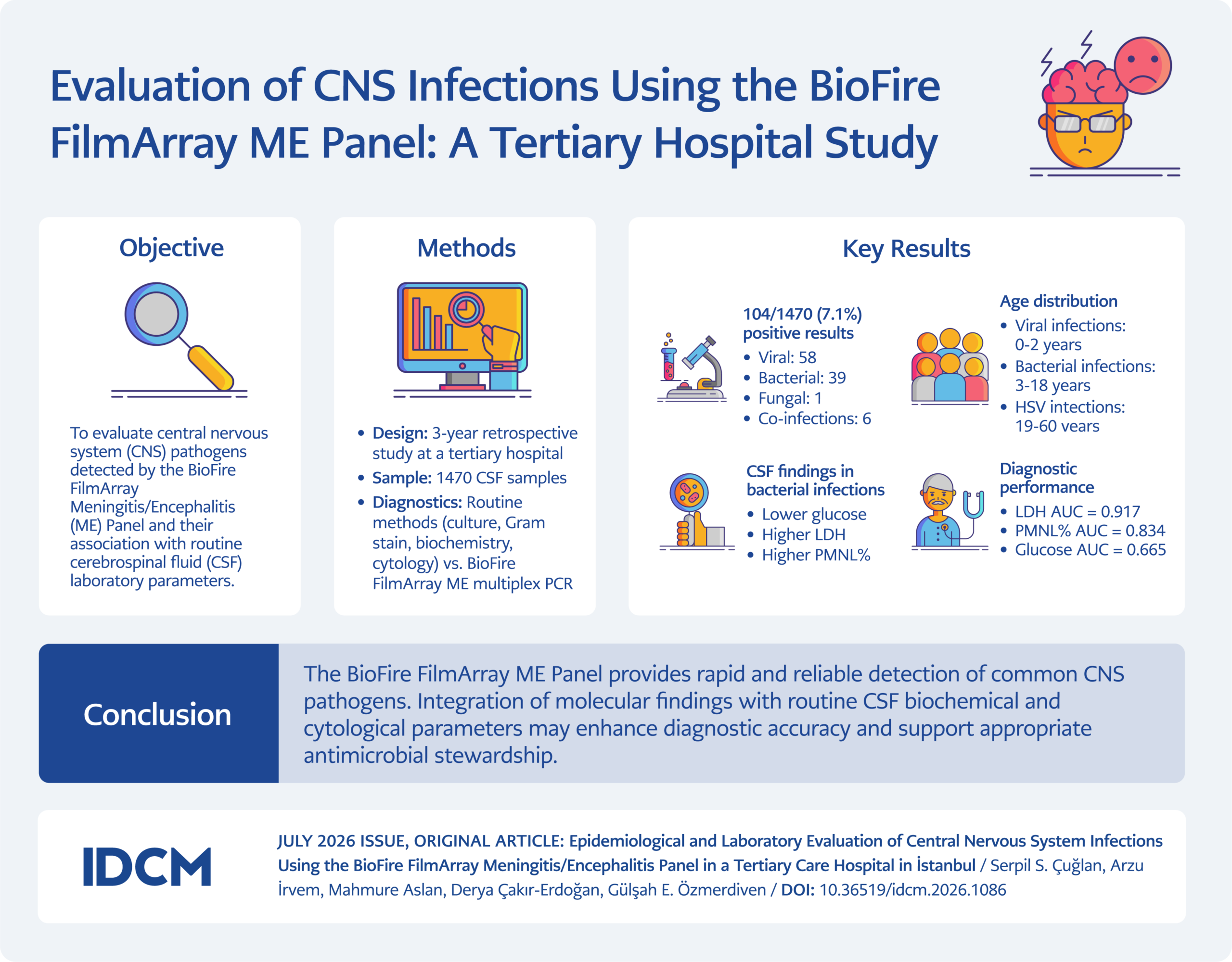
Graphical Abstract
Introduction
Acute bacterial meningitis (ABM) is a rapidly progressive disease with outbreak potential and remains associated with substantial morbidity and mortality worldwide (1). Although vaccination programs have significantly reduced the incidence of several major pathogens, ABM continues to pose a significant public health challenge. Streptococcus pneumoniae, Neisseria meningitidis, and Haemophilus influenzae type b are among the most common causative agents of bacterial meningitis (2). However, the epidemiology of meningitis varies considerably according to age distribution, geographic region, seasonal factors, population immunity, socioeconomic conditions, and local endemic patterns (3).
Viral infections of the central nervous system (CNS) present with a wide spectrum of clinical manifestations, including meningitis, encephalitis, post-infectious encephalomyelitis, and chronic or slowly progressive neurological syndromes. Viral meningitis is generally self-limiting, whereas viral encephalitis, most commonly caused by herpes simplex virus (HSV), may result in significant neurological sequelae if not promptly diagnosed and treated (4). Chronic meningitis, defined as symptoms persisting for more than four weeks, is most commonly associated with tuberculosis and fungal infections.
The diagnosis of CNS infections is often challenging due to overlapping clinical features of bacterial and viral etiologies. Conventional diagnostic methods, including culture and serologic testing, may be time-consuming and lack sensitivity, particularly when empirical antimicrobial therapy is initiated prior to cerebrospinal fluid (CSF) sampling. Molecular diagnostic techniques have markedly improved the rapid identification of causative pathogens. Enteroviruses, parechoviruses, respiratory viruses (including SARS-CoV-2, metapneumovirus, measles, and mumps), and herpes viruses (HSV-1, HSV-2, human herpesvirus 6 (HHV-6), and varicella-zoster virus [VZV]) are well-recognized causes of endemic or epidemic CNS infections (5).
The BioFire FilmArray Meningitis/Encephalitis (ME) Panel is a multiplex polymerase chain reaction (PCR) assay that enables rapid, simultaneous detection of 14 common CNS pathogens directly from CSF samples. The availability of such molecular platforms has facilitated early diagnosis, optimized antimicrobial therapy, and improved cost-effectiveness in the management of suspected meningitis and encephalitis. This study aimed to evaluate the performance of the BioFire FilmArray ME Panel compared with conventional CSF analysis.
Materials and Methods
This study included cerebrospinal fluid samples analyzed at a tertiary care hospital laboratory over a three-year post-pandemic period (2021–2023). Cell counts, culture results, and biochemical parameters (glucose, chloride, lactate dehydrogenase, sodium, potassium, and total protein) of patients with positive CSF PCR results were retrospectively reviewed and recorded. Patient demographic characteristics, such as age, sex, and the season during which the causative agent was identified, were also recorded. Duplicate samples from the same patient were excluded from the study.
Multiplex PCR Panel
Cerebrospinal fluid samples were analyzed using the FilmArray multiplex PCR system (BioFire FilmArray ME Panel; bioMérieux, Marcy-l’Étoile, France) in accordance with the manufacturer’s instructions. The assay performs automated nucleic acid extraction, reverse transcription, and nucleic acid amplification for 14 pathogens, including bacteria (Escherichia coli K1, Haemophilus influenzae, Listeria monocytogenes, Neisseria meningitidis, Streptococcus agalactiae, and Streptococcus pneumoniae); viruses [cytomegalovirus (CMV), enterovirus, HSV-1, HSV-2, HHV-6, human parechovirus (HPeV), and VZV]; and yeast [Cryptococcus (Cryptococcus neoformans/Cryptococcus gattii)] with results available within approximately one hour. After completion of the two control runs included in each FilmArray kit [targeting an RNA transcript from Schizosaccharomyces pombe and a DNA target], the FilmArray software automatically generated the results.
Bacterial Culture
All samples included in the study were processed for cell counting, culture, and Gram staining. Cerebrospinal fluid samples were inoculated onto sheep blood agar, MacConkey agar, chocolate agar, Sabouraud dextrose agar, and chromogenic agar according to standard procedures. The inoculated plates were incubated at 35–37°C in 5% CO2. Plates were examined for microbial growth every 24 hours. Samples showing no growth after 72 hours were considered culture negative. When growth was detected, isolates were identified by matrix-assisted laser desorption ionization time-of-flight mass spectrometry (MALDI-TOF MS; Bruker Daltonics, Billerica, MA, USA) analysis. Antimicrobial susceptibility testing was performed using the VITEK 2 system (bioMérieux, Marcy-l’Étoile, France).
Biochemical Analysis
Biochemical analyses and cell counts were performed on all samples included in the study. These analyses were performed using methods standardized according to the International Federation of Clinical Chemistry and Laboratory Medicine (IFCC). Cerebrospinal fluid protein levels were measured using a turbidimetric assay, CSF glucose levels using the hexokinase reference method, and CSF lactate dehydrogenase (LDH) levels using a spectrophotometric assay (Cobas 6000 c501 autoanalyzer; Roche Diagnostics, Mannheim, Germany). Sodium, potassium, and chloride levels were measured using the indirect ion-selective electrode (ISE) method (Cobas 6000 c501 autoanalyzer; Roche Diagnostics, Mannheim, Germany). Cerebrospinal fluid leukocyte counts were determined using both the impedance method (BC-6800 Plus hematology analyzer; Mindray, China) and a Nageotte counting chamber.
Statistical Analysis
All statistical analyses were performed using the IBM SPSS Statistics for Windows, version 22.0 (IBM Corp., Armonk, NY, USA). Categorical variables were expressed as counts and percentages and compared between groups using the chi-square test. Continuous variables were expressed as mean ± standard deviation (SD) when normally distributed. Data normality was assessed using the Kolmogorov-Smirnov test. According to the results of this analysis, the independent-samples t-test was applied to normally distributed continuous variables. For non-normally distributed continuous variables, the Mann-Whitney U test was used, and the results were expressed as medians. Area under the curve (AUC) values were estimated using receiver operating characteristic (ROC) analysis, and 95% confidence intervals (CIs) were calculated by bootstrap resampling.
Results
A total of 1470 CSF samples were analyzed in our laboratory between 2021 and 2023. Pathogens were detected in 104 samples (7.1%), including 57 viral, 39 bacterial, one fungal, and six co-infections. Cryptococcus neoformans was detected in one 24-year-old male patient and was excluded from comparative statistical analyses because it occurred as a single case.
On culture, S. pneumoniae growth was detected in one sample, while no bacterial growth was observed in the remaining 38 samples that were identified by multiplex PCR as containing bacterial pathogens. Cryptococcus neoformans was also isolated from the corresponding PCR-positive fungal case. Culture findings were concordant with PCR results in these cases.
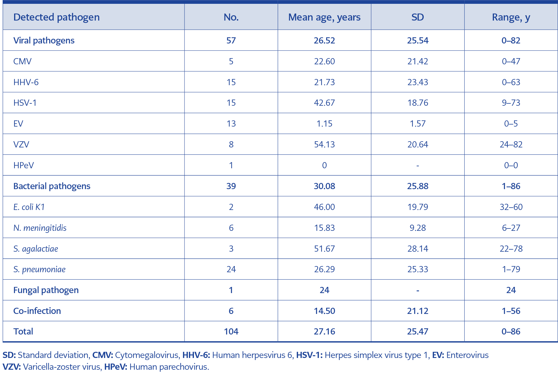
Table 1. Age distribution of patients according to detected pathogens.
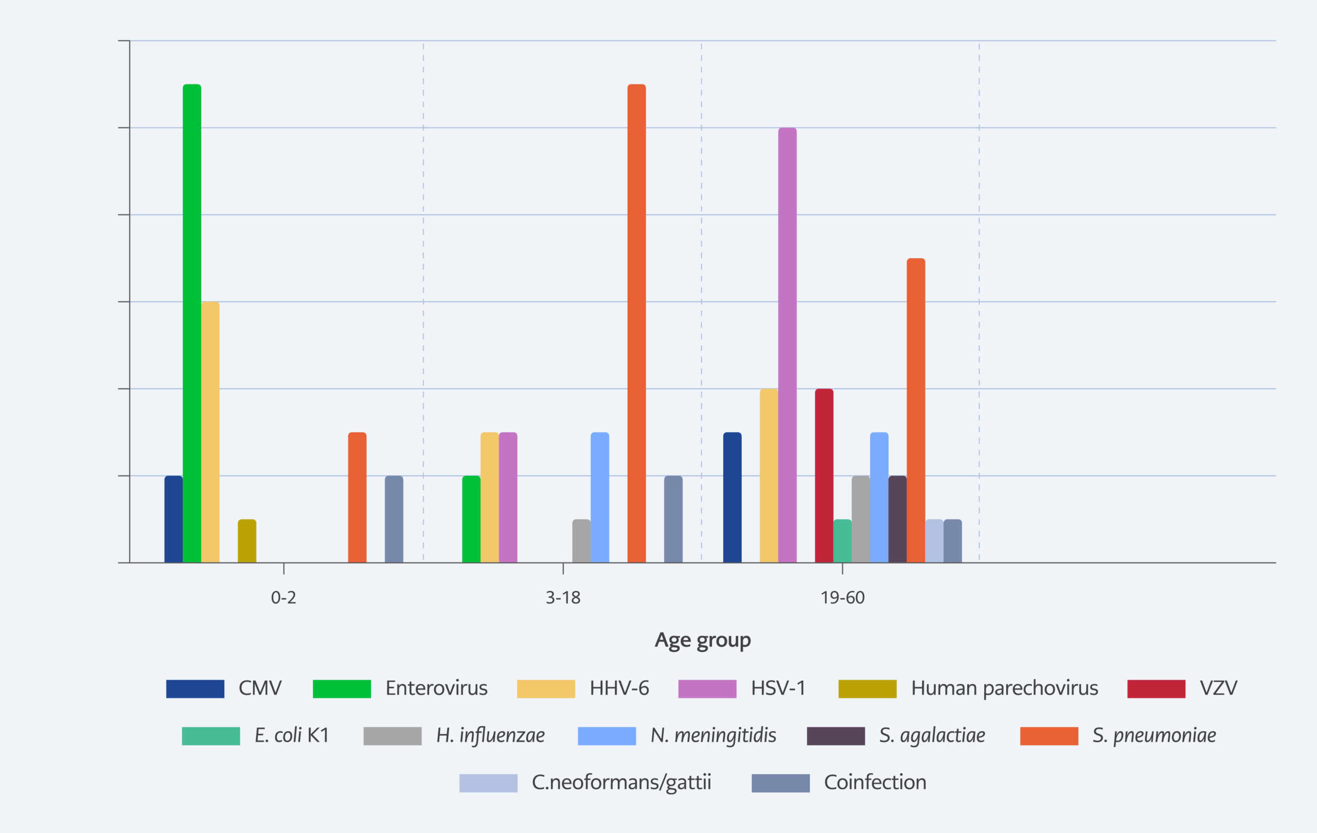
Figure 1. Distribution of detected pathogens according to age groups.
The patients ranged in age from 0 to 86 years (mean ± SD, 27.16 ± 25.47 years; median, 21 years). The distribution of pathogens by age group is presented in Table 1 and Figure 1. Viral infections were significantly more frequent among children aged 0–2 years (p = 0.002), whereas bacterial infections were significantly more common in patients aged 3–18 years (p = 0.027). Additionally, viral infections, particularly HSV, were significantly more prevalent among patients aged 19–60 years (p = 0.008). No statistically significant association was found between sex and pathogen distribution (p = 0.185).
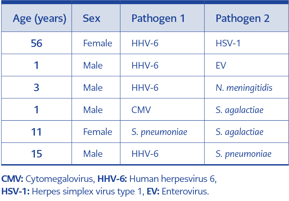
Table 2. Polymicrobial detections identified by the BioFire FilmArray ME Panel.
Six co-infections were identified by the BioFire FilmArray ME Panel. The detected pathogen combinations are presented in Table 2.
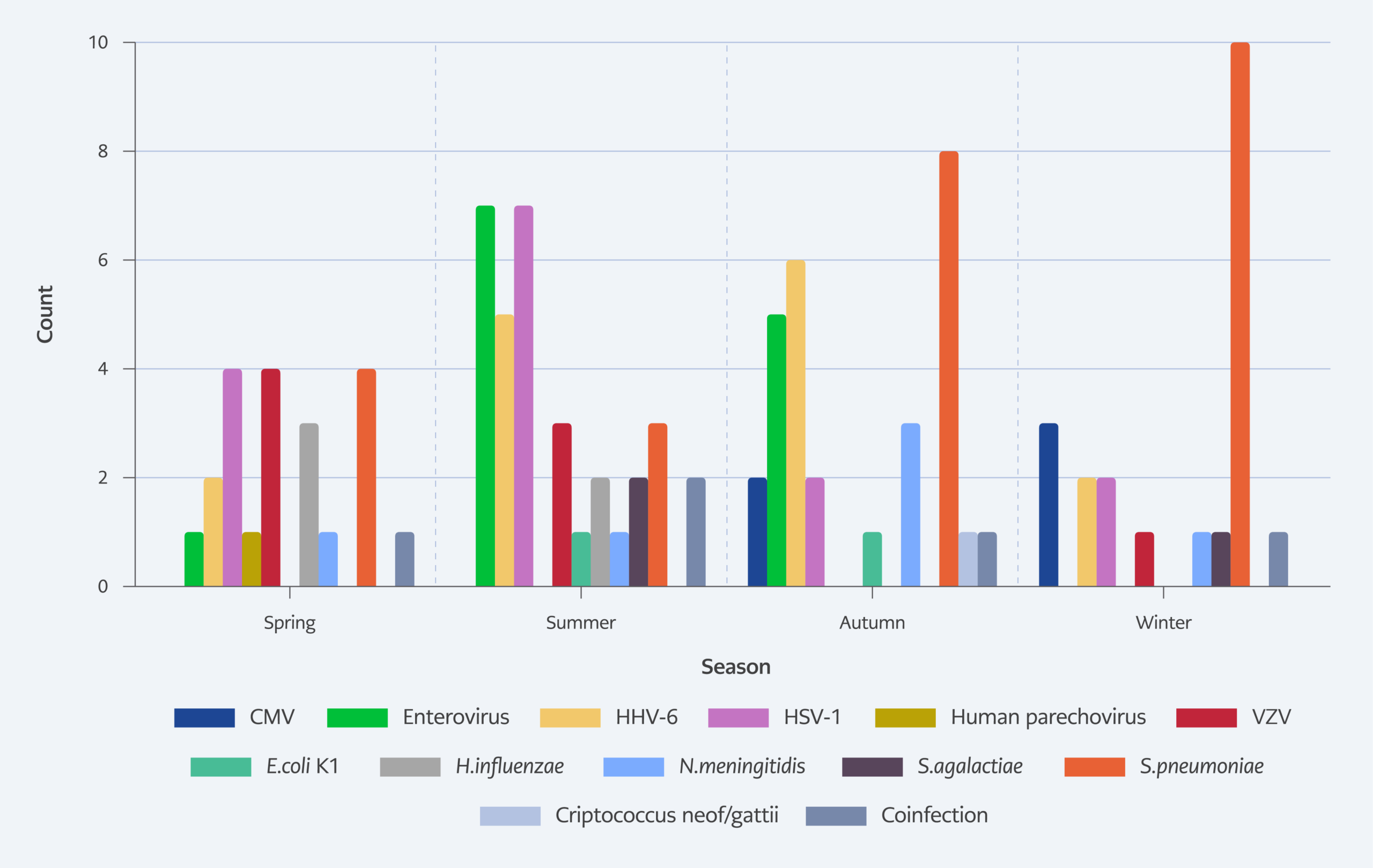
Figure 2. Seasonal distribution of detected pathogens.
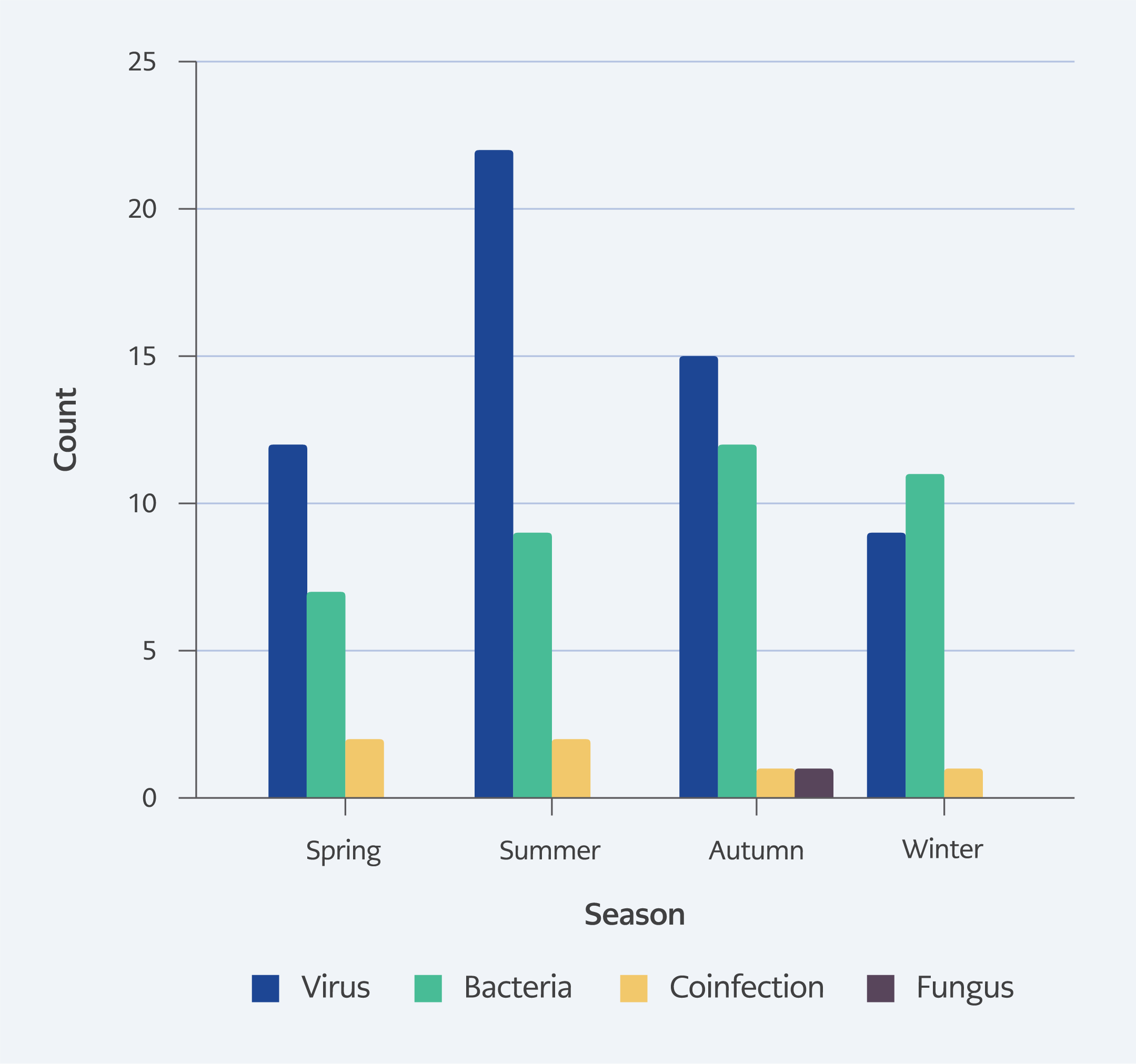
Figure 3. Seasonal distribution of pathogen groups.
The seasonal distribution of pathogens is presented in Figures 2 and 3. Although viral infections appeared more frequent during the summer months, no statistically significant association with season was observed (p = 0.293).
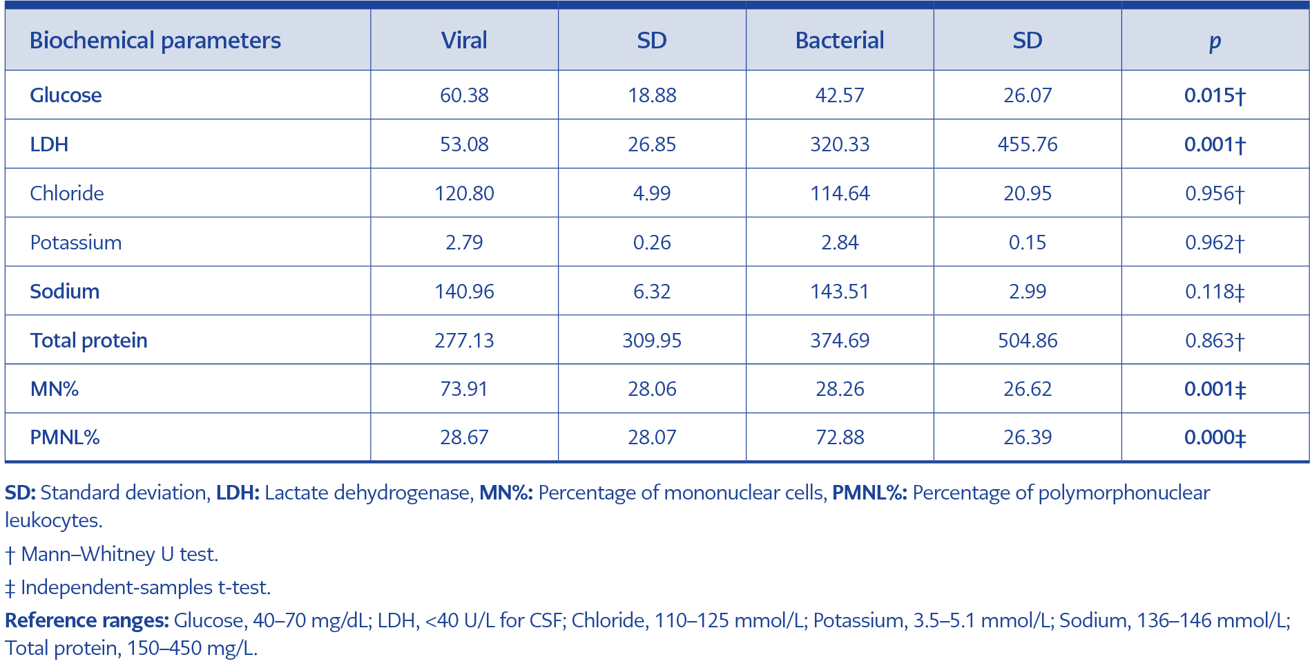
Table 3. Comparison of cerebrospinal fluid biochemical parameters between viral and bacterial infections.
The distribution of CSF biochemical parameters according to viral and bacterial etiologies is shown in Table 3. The mean CSF glucose level was significantly lower in the bacterial group than in the viral group (42.57 ± 26.07 mg/dL vs. 60.38 ± 18.88 mg/dL; p = 0.015). Conversely, CSF LDH levels were significantly higher in bacterial cases (320.33 ± 455.76 IU/L) than in viral cases (53.08 ± 26.85 IU/L; p = 0.001). No statistically significant differences were observed between groups in chloride, sodium, potassium, or total protein levels (Table 3).
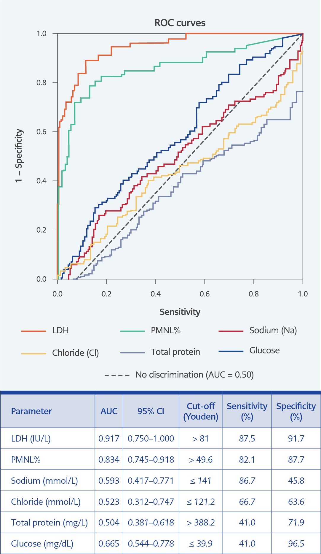
Figure 4. ROC analysis of CSF laboratory parameters for differentiating bacterial and viral etiologies.
Receiver operating characteristic analysis evaluated the performance of CSF glucose, LDH, PMNL%, sodium, chloride, and total protein levels in differentiating bacterial from viral CNS infections. Bacterial infection was defined as the positive outcome. Lactate dehydrogenase demonstrated the highest discriminatory performance (AUC = 0.917; 95% CI, 0.750–1.000), followed by PMNL percentage (AUC = 0.834; 95% CI, 0.745–0.918). Cerebrospinal fluid glucose demonstrated moderate discriminatory ability (AUC = 0.665; 95% CI, 0.544–0.778), whereas sodium, chloride, and total protein showed limited discriminatory performance. Optimal cutoff values, sensitivity, and specificity were determined using the Youden index. The diagonal line represents the line of no discrimination (Figure 4).
Discussion
The diagnosis of viral meningitis and encephalitis remains challenging due to overlapping clinical features and limitations of conventional diagnostic methods. Since its approval by the U.S. Food and Drug Administration (FDA) in 2015, the BioFire FilmArray ME Panel has been increasingly adopted worldwide as a rapid multiplex PCR assay capable of detecting common CNS pathogens directly from CSF samples (6).
Despite its advantages, concerns regarding false-positive and false-negative results have been reported, particularly for S. pneumoniae and group B Streptococcus, as well as false-negative HSV results and detection of latent viral infections such as CMV and HHV-6 (7,8,9). In our study, PCR-positive results were not routinely confirmed by alternative molecular methods. However, culture growth was observed in one S. pneumoniae case and in the single C. neoformans case, demonstrating concordance between PCR and conventional microbiological methods.
A notable finding of this study was the high proportion of culture-negative but PCR-positive bacterial cases. While prior antibiotic exposure is a well-recognized factor that reduces culture sensitivity, detailed antibiotic histories were not consistently available because of the retrospective design and the centralized laboratory serving multiple hospitals (10). In addition, other factors, such as low bacterial load, fastidious organisms, and pre-analytical variables, may have contributed to culture negativity. These findings emphasize the complementary role of molecular diagnostics, particularly in settings where conventional culture methods have limited sensitivity.
Rapid laboratory reporting of HSV results plays a critical role in clinical management. Early exclusion of HSV infection may prevent unnecessary or prolonged empirical acyclovir therapy and its associated nephrotoxicity, particularly in pediatric patients (10,11). Similarly, rapid identification of viral etiologies such as enterovirus or Epstein-Barr virus (EBV) may reduce inappropriate antibiotic use in cases of aseptic meningitis (12). The BioFire FilmArray ME Panel is also the FDA-approved assay for detecting human parechovirus (HPeV), an important cause of meningitis in infants (13).
Interpretation of positive results for certain viral targets, such as HHV-6 and CMV, requires careful clinical correlation, as these detections may represent latent infection or viral reactivation rather than active CNS disease (14). In our cohort, several such cases could not be confirmed by additional diagnostic methods, further highlighting the importance of integrating laboratory findings with the patient’s clinical presentation.
Routine CSF cytological and biochemical parameters demonstrated significant differences between bacterial and viral infections. Cerebrospinal fluid glucose levels were lower, whereas LDH levels and PMNL percentages were higher in bacterial cases. ROC analysis demonstrated high discriminatory performance for LDH and PMNL%, supporting their potential utility as adjunctive markers for differentiating bacterial from viral etiologies. Co-infections were observed in a limited number of cases. The clinical significance of these findings remains uncertain, and they may represent true co-infection, colonization, or detection of latent viral DNA. Therefore, such results should be interpreted cautiously in the appropriate clinical context.
The patient’s immune status should always be considered when interpreting ME Panel results. Traumatic lumbar puncture may lead to peripheral blood contamination and potentially misleading results (7). Additionally, the absence of Mycobacterium tuberculosis targets in the BioFire FilmArray ME Panel represents a limitation in regions where tuberculous meningitis remains prevalent. Empirical treatment is often initiated in suspected tuberculous meningitis because of the low sensitivity and prolonged turnaround time of conventional diagnostic methods.
Cost analyses have suggested that although the per-test cost of the ME Panel is relatively high, overall healthcare expenditure may not increase significantly when molecular testing is integrated into routine diagnostics, owing to reductions in unnecessary antimicrobial use and length of hospital stay (15).
The epidemiology of bacterial meningitis varies according to age and geographic region (16–18). In our study, bacterial pathogens were detected in 39 (2.6%) and viral pathogens in 58 (3.9%) CSF samples, with an additional six co-infections. Viral infections predominated in children aged 0–2 years, whereas bacterial infections were more common among older children. The relatively low incidence of neonatal bacterial meningitis in our cohort may reflect effective vaccination programs and improved perinatal healthcare practices (19–21).
This study has several limitations. Mycobacterium tuberculosis was not included because it is not covered by the PCR panel used in this study. PCR-positive findings were not systematically confirmed using additional diagnostic methods. Biochemical parameters of PCR-negative samples were within or close to the reference ranges and were therefore excluded from statistical evaluation because these samples may have represented non-infectious neurological conditions rather than CNS infections. In addition, because the study was conducted in a centralized laboratory serving 12 affiliated hospitals, detailed clinical follow-up data and information on prior antibiotic use were not consistently available.
In conclusion, the BioFire FilmArray ME Panel provides rapid and reliable detection of common CNS pathogens and serves as a valuable adjunct to routine CSF laboratory analysis. When interpreted together with CSF cytological and biochemical parameters, molecular results may improve diagnostic accuracy and support appropriate antimicrobial stewardship. The predominance of viral etiologies in infants and the relatively low incidence of neonatal bacterial meningitis in our cohort may reflect effective vaccination strategies and improvements in maternal healthcare practices.