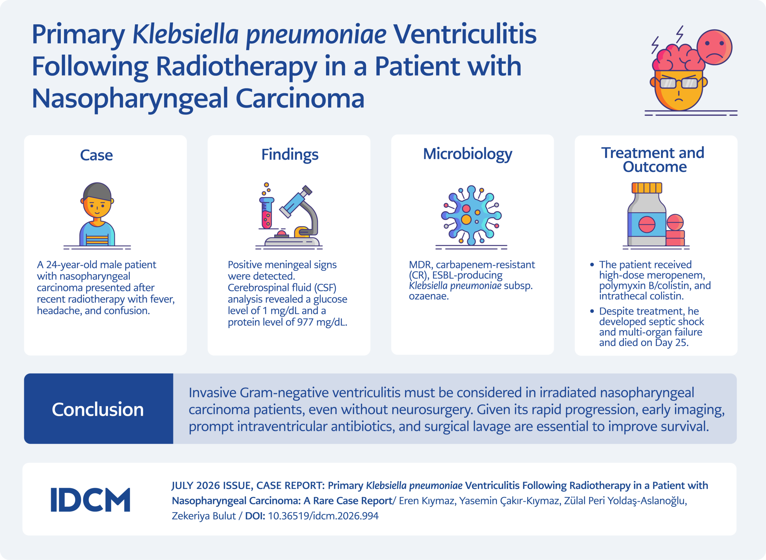
Graphical Abstract
Introduction
Bacterial meningitis and ventriculitis are severe central nervous system (CNS) infections characterized by high morbidity and mortality, particularly in immunocompromised patients (1). In clinical practice, ventriculitis is predominantly encountered as a secondary complication of invasive neurosurgical procedures, such as the placement of external ventricular drains (EVDs) or following intraventricular hemorrhage (2). Conversely, primary ventriculitis is defined as an infection of the ventricular system occurring via hematogenous spread or contiguous extension in the absence of prior surgical intervention and is exceedingly rare in the literature (3). This rarity poses a significant diagnostic challenge for clinicians, often leading to delayed recognition and management.
Gram-negative bacteria, particularly multidrug-resistant (MDR) Klebsiella pneumoniae strains, are associated with limited therapeutic options and mortality rates reaching up to 70% in CNS infections (4,5). Patients with malignancies receiving radiotherapy are at an elevated risk for the translocation of such aggressive pathogens due to immune dysfunction, disruption of anatomical barriers, and mucosal damage. The development of primary K. pneumoniae ventriculitis complicated by acute hydrocephalus in a patient with nasopharyngeal carcinoma without a history of neurosurgery represents an unusual clinical presentation.
In this report, we present a case of primary K. pneumoniae ventriculitis in a young patient with nasopharyngeal carcinoma following radiotherapy. Despite aggressive interventions, including intrathecal colistin and surgical drainage, the clinical course was fatal. This case aims to highlight the aggressive nature of primary Gram-negative ventriculitis and to contribute to the limited literature by emphasizing its diagnostic pitfalls and management complexities.
Case
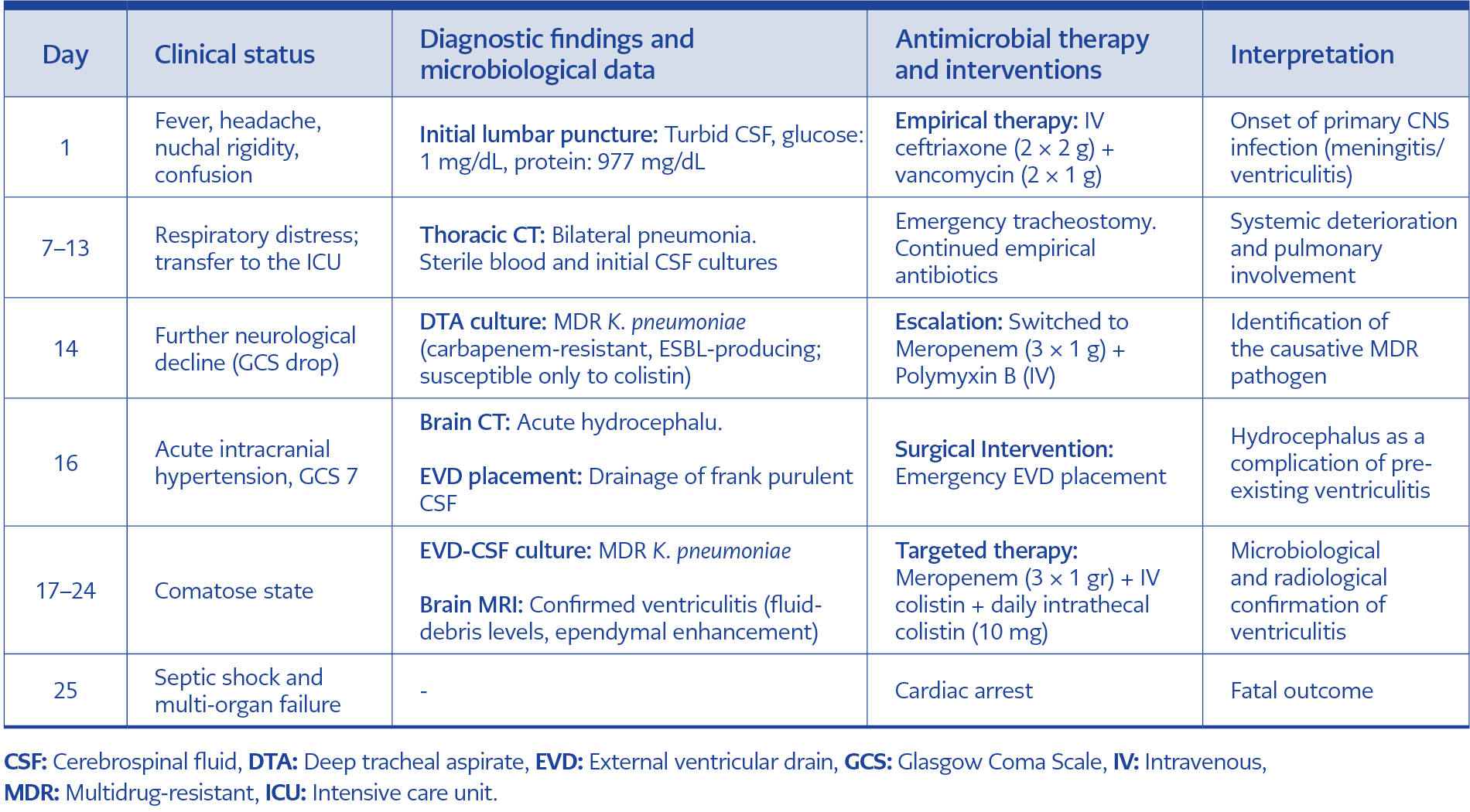
Table 1. Clinical progression, diagnostic findings, and antimicrobial management timeline.
A 24-year-old male, who had been followed for six years with a diagnosis of nasopharyngeal carcinoma, presented to the emergency department with fever and headache after completing a 10-day course of radiotherapy two months earlier. The chronological progression of the patient’s clinical status, diagnostic findings, and antimicrobial therapy is summarized in Table 1. Upon physical examination, his general condition was fair to poor; he was confused, with limited orientation and cooperation. Vital signs were as follows: temperature, 38.2°C; blood pressure, 110/60 mmHg; heart rate, 94 bpm; and SpO2, 98%. Neurological examination revealed nuchal rigidity and positive Kernig and Brudzinski signs.
A lumbar puncture was performed, yielding turbid CSF. CSF biochemistry showed profound hypoglycorrhachia, with a glucose level of 1 mg/dL (concomitant blood glucose, 110 mg/dL) and an elevated protein level of 977 mg/dL. Although initial Gram stain and culture results were negative, these findings were interpreted as early evidence of a severe pyogenic process likely already involving the ventricular system (7). Empirical therapy was initiated with intravenous (IV) ceftriaxone (2 × 2 g) and vancomycin (2 × 1 g).
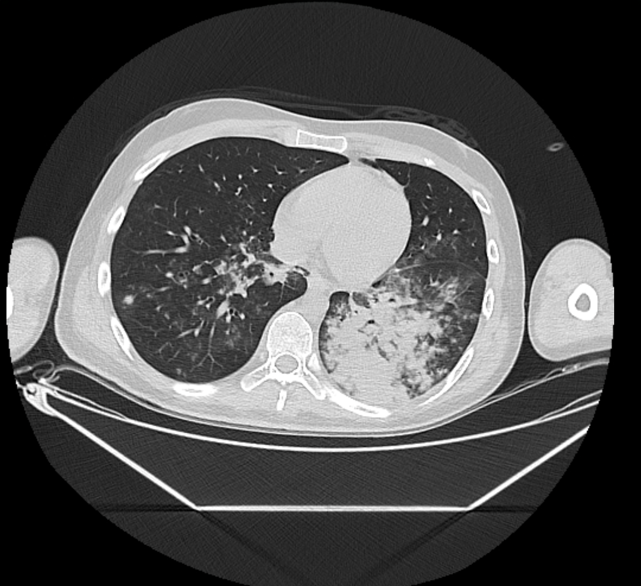
Figure 1. Thoracic CT scan showing bilateral ground-glass opacities in the upper zones and consolidation in the left middle zone, interpreted as pneumonia.
During follow-up, the patient developed respiratory distress. Chest computed tomography (CT) demonstrated bilateral ground-glass opacities in the upper zones and pneumonic consolidations in the left middle zone, consistent with pneumonia (Figure 1). Initial blood, CSF, and sputum cultures obtained during the first week remained sterile. Due to clinical deterioration and a declining Glasgow Coma Scale (GCS) score, the patient was transferred to the intensive care unit (ICU). Following unsuccessful intubation attempts, an emergency tracheostomy was performed to secure the airway.
On Day 14, following further clinical deterioration, K. pneumoniae was isolated from deep tracheal aspirate (DTA) cultures. Microbiological identification was performed using matrix-assisted laser desorption/ionization time-of-flight mass spectrometry (MALDI-TOF MS) with the Bruker IVD MALDI Biotyper 2.3 system (Bruker Daltonik GmbH, Bremen, Germany). Antimicrobial susceptibility testing was conducted using the fully automated BD Phoenix 100 system (Becton Dickinson, Sparks, MD, USA) and interpreted in accordance with European Committee on Antimicrobial Susceptibility Testing (EUCAST) guidelines. Extended-spectrum beta-lactamase (ESBL) positivity was detected by the double-disk synergy test. Notably, colistin susceptibility was specifically determined using the broth microdilution method. The isolate was categorized as MDR based on the criteria established by Magiorakos et al. (8), exhibiting a carbapenem-resistant and ESBL-producing phenotype. Consequently, the antimicrobial therapy was switched to IV meropenem (3 × 1 g/day) and polymyxin B (loading dose of 25,000 IU/kg, followed by a maintenance dose of 12,500 IU/kg twice daily [BID]).
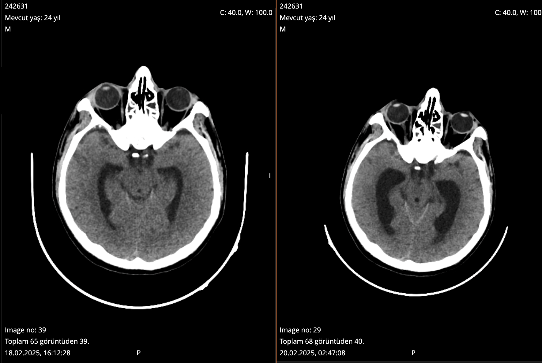
Figure 2. Non-contrast brain CT scan demonstrating acute hydrocephalus with significant ventricular enlargement following neurological deterioration.
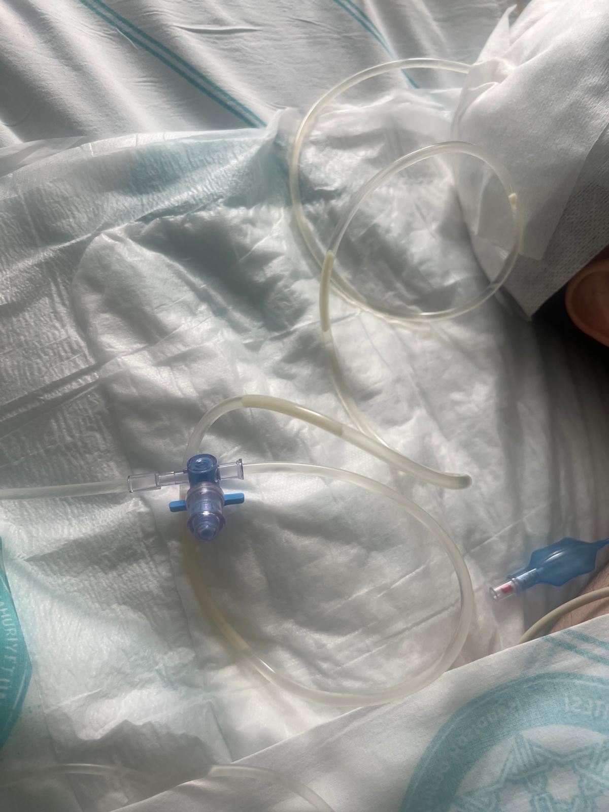
Figure 3. Gross appearance of purulent CSF drainage observed during the emergency placement of an EVD.
Following further neurological decline on Day 16, a brain CT revealed acute hydrocephalus (Figure 2), necessitating the emergency placement of an EVD, which yielded purulent CSF (Figure 3). On Day 17, K. pneumoniae was subsequently isolated from the CSF. Notably, this isolate was also identified as the rare subspecies K. pneumoniae ssp. ozaenae DSM 16358T with high-confidence MALDI-TOF MS scores (2.425 for DTA and 2.160 for CSF) and exhibited an identical antibiotic susceptibility profile to the DTA isolate. Although genetic characterization for clonality is not routinely performed in our clinical practice, the isolation of the same rare biotype with an identical resistance profile from both sites, coupled with temporal consistency, strongly suggested a hematogenous translocation from the respiratory tract to the central nervous system.
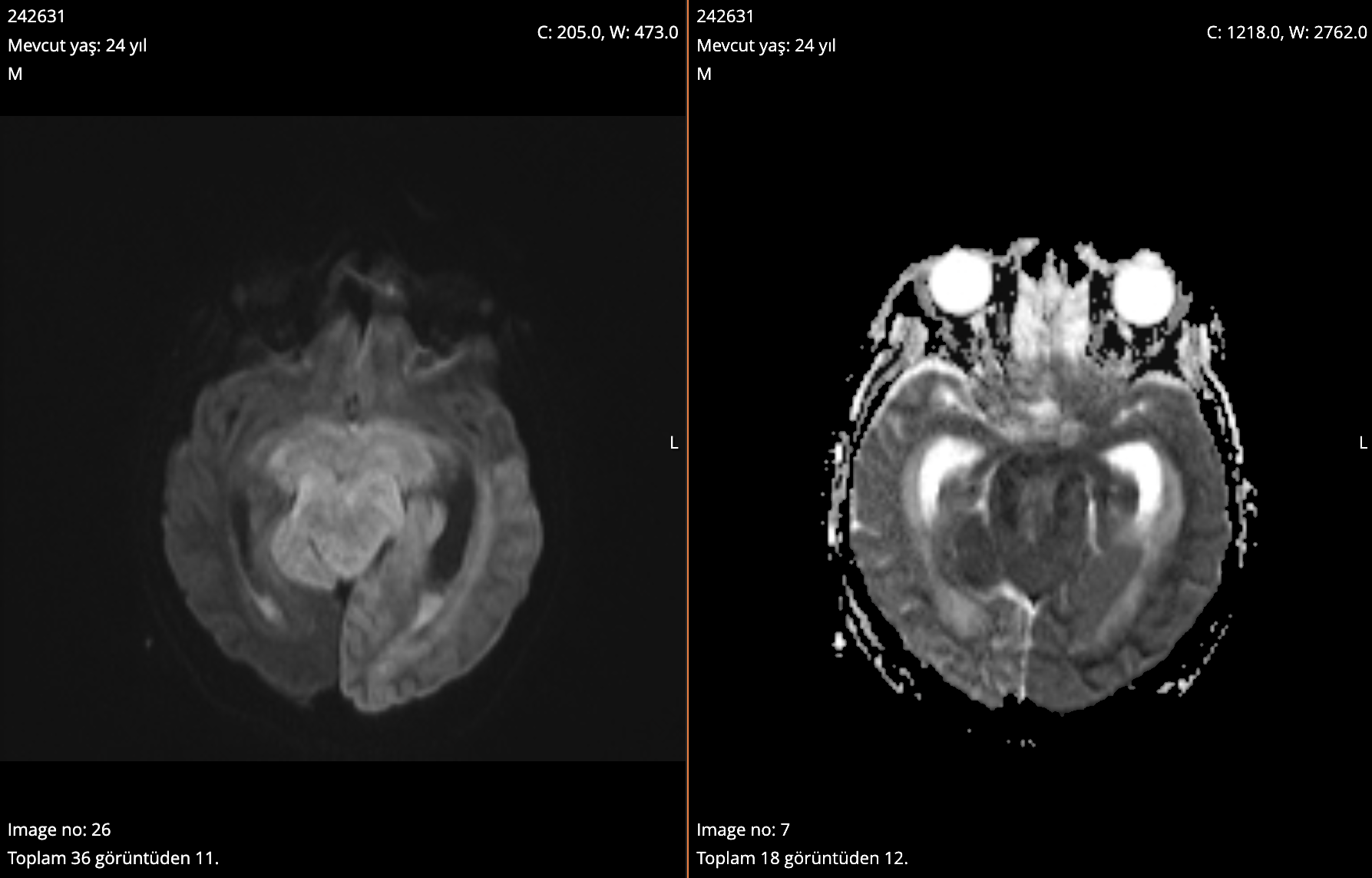
Figure 4. Contrast-enhanced brain MRI demonstrating fluid-debris levels within the occipital horns of the lateral ventricles and restricted diffusion, consistent with pyogenic ventriculitis.
Contrast-enhanced brain magnetic resonance imaging confirmed ventriculitis with fluid-debris levels and restricted diffusion (Figure 4). Considering the poor CSF penetration of systemic polymyxins, the regimen was intensified as a salvage strategy with high-dose meropenem (3 × 2 g), systemic colistin, and daily intrathecal colistin (10 mg) to achieve bactericidal concentrations within the ventricular system (4). Despite these intensive measures, the patient progressed to septic shock and multi-organ failure, leading to a fatal outcome on Day 25.
Discussion
Primary ventriculitis is generally defined as a spontaneous infection of the ventricular system occurring in the absence of prior neurosurgical intervention or trauma (1,2). It can rarely manifest as primary ventriculitis in patients without a surgical history (3). The interpretation of this case as primary ventriculitis remains valid despite the isolation of K. pneumoniae from the DTA three days prior to its isolation from the ventricular CSF. In neurosurgical terminology, secondary ventriculitis is fundamentally defined as an iatrogenic infection following invasive procedures, such as EVD placement or craniotomy (4). In contrast, primary ventriculitis occurs spontaneously via hematogenous or contiguous spread in the absence of surgical intervention. Since our patient had no prior neurosurgical history, the pulmonary involvement likely represented a concurrent focus or the hematogenous source, which is a recognized pathway for primary infection. Furthermore, the profound hypoglycorrhachia (1 mg/dL) and meningeal signs present on Day 1 provide strong evidence that the CNS infection was established at admission, well before the clinical progression of pneumonia or any surgical breach. Therefore, the chronological sequence of positive cultures does not exclude the diagnosis of primary ventriculitis, as the infection arose spontaneously rather than as a procedural complication.
The microbiological findings obtained later in the clinical course support this suspected pathogenesis. Specifically, K. pneumoniae was isolated from the DTA on Day 14 and subsequently from the ventricular CSF on Day 17. The isolation of the same rare subspecies with an identical resistance profile from both sites, coupled with temporal consistency, strongly suggests hematogenous spread from the respiratory tract to the central nervous system, although molecular analysis was not performed to confirm clonality. Furthermore, the aggressive nature of the infection and its rapid progression to ventricular empyema in a non-neurosurgical patient raise the suspicion of a hypervirulent K. pneumoniae (hvKp) phenotype. hvKp strains are characterized by increased capsular polysaccharide production and specific virulence genes (such as rmpA and iucA), which enhance their ability to cause invasive disease, including primary CNS infections, by translocating through mucosal barriers, a process likely facilitated in this patient by radiotherapy-induced tissue damage (6). However, because phenotypic or molecular investigations for hypervirulence markers are not performed as part of routine microbiology practice at our institution, the hypervirulence status of the isolated strain could not be further evaluated.
Radiological imaging is vital for the diagnosis of ventriculitis. Ventricular dilatation, ependymal enhancement, and fluid-debris levels observed in our patient are characteristic findings (7,9). Diffusion-weighted imaging is particularly sensitive, as purulent exudates typically exhibit diffusion restriction, providing a diagnostic advantage over conventional sequences (10). In this case, the rapid progression and heavy debris accumulation characteristic of invasive K. pneumoniae ventriculitis were clearly delineated by these imaging modalities (10,11).
The management of MDR Gram-negative ventriculitis is severely limited by the pharmacokinetics of systemic antibiotics. In the present case, the escalation of antimicrobial therapy followed a stepwise rationale based on microbiological findings and pharmacokinetic challenges. Initially, upon the isolation of MDR K. pneumoniae from the respiratory tract, the regimen was switched to IV meropenem (3 × 1 g/day) and polymyxin B (loading dose of 25,000 IU/kg, followed by a maintenance dose of 12,500 IU/kg BID). Following the subsequent confirmation of ventriculitis by culture of EVD-derived CSF, the treatment was further intensified by doubling the meropenem dosage to 2 g every 8 hours to maximize peak concentrations. Simultaneously, daily intrathecal colistin (10 mg) was initiated as a salvage strategy. This decision aligns with the 2017 Infectious Diseases Society of America (IDSA) guidelines, which recommend localized antimicrobial delivery for MDR Gram-negative infections when systemic penetration is inadequate or clinical response is poor (4). Systemic polymyxins exhibit particularly poor blood-brain barrier penetration even in the presence of inflammation, necessitating direct intraventricular administration to achieve therapeutic bactericidal levels (12–14). However, the management of such cases is often complicated by dense purulent material and intraventricular flocculation, which can create adhesions and hinder antibiotic bioavailability. While some centers advocate for early ventricular lavage to reduce this purulent burden (15), our patient progressed to septic shock and multi-organ failure despite emergency surgical drainage and intensive antimicrobial therapy.
The fatal outcome in this case was likely multifactorial. The suppressive effect of radiotherapy on both innate and adaptive immunity, combined with the loss of mucosal barrier integrity, may have rendered the patient more susceptible to invasive pathogens. Furthermore, prolonged hospitalization, ICU stay, and invasive airway management likely served as catalysts for K. pneumoniae colonization and subsequent systemic dissemination.
Conclusion
The risk of invasive Gram-negative ventriculitis must be considered in patients with nasopharyngeal carcinoma and a history of radiotherapy, even in the absence of neurosurgical intervention. Given the rapid progression of primary Gram-negative ventriculitis, a multidisciplinary approach incorporating early radiological assessment, prompt intraventricular antibiotic therapy, and early surgical lavage appears to be the only viable strategy for improving survival.