Introduction
Diabetes mellitus (DM) is a major global health burden, and its prevalence has nearly doubled over the last 25 years. Individuals with DM are significantly more prone to developing skin and soft tissue infections (SSTIs), including cellulitis, osteomyelitis, and post-operative wound infections (1). In a large retrospective analysis of more than two million patients with SSTIs, 10% were diabetic (2). Among outpatients, the rate of SSTI-related complications was more than five times higher in individuals with DM than in those without diabetes (2).
Diabetic hand infections (DHIs) represent a particularly serious form of SSTI, as they can progress rapidly and may lead to permanent functional impairment or amputation. Although relatively uncommon, DHIs are reported worldwide and appear more frequently in tropical regions (3,4). Even minor trauma may trigger infection, as hyperglycemia impairs neutrophil function and immune defense, while microangiopathy and neuropathy delay healing. The interplay of these factors collectively creates a favorable environment for fungal colonization and persistence (1,5–7).
Here, we describe a case of DHI caused by Candida tropicalis that required repeated surgical debridement, in addition to systemic antifungal therapy.
Case
A 62-year-old woman with a history of type 2 DM, hypertension, and coronary artery disease presented with a 4-day history of redness, swelling, and severe pain in the left thumb. She reported a cactus-torn injury to the distal left thumb. At an outside facility, she had received intravenous ampicillin-sulbactam (8 g/day) for presumed bacterial infection, but her symptoms did not improve. She was referred to our department with suspected DHI and a persistent lesion of 14 days duration.

Figure 1. An edematous and erythematous lesion with purulent formation in the subcutaneous area.
Physical examination revealed a swollen, erythematous thumb with purulent drainage from a subcutaneous collection (Figure 1). Laboratory tests revealed a white blood cell count of 6500/μL with 70% neutrophils, an elevated C-reactive protein level of 15 mg/L (reference: 0–5), and an erythrocyte sedimentation rate of 30 mm/hour. Chest x-ray was unremarkable, and a tuberculin skin test with purified protein derivative showed no induration. Urinalysis, basic chemistry, and hepatic function results were within normal ranges. The patient was afebrile.
Upon admission, the patient was consulted by a plastic surgeon for evaluation and management of necrotizing cellulitis and purulent discharge. Emergency surgical debridement was performed, and tissue samples were sent for laboratory analysis. Direct microscopy, culture, and molecular testing were conducted. Sabouraud dextrose agar yielded creamy, smooth colonies after 48 hours of incubation at 37°C. Microscopic examination with lactophenol cotton blue staining demonstrated yeast cells with pseudohyphae, consistent with Candida spp. The isolate was identified as C. tropicalis using the VITEK® 2 automated system (bioMérieux, Marcy-l’Étoile, France). No additional fungal growth was detected in the culture following four weeks of incubation at both 35°C and room temperature.
Given the progressive swelling and emerging necrosis, empiric piperacillin-tazobactam and vancomycin were initiated; however, no clinical improvement was observed. Antifungal susceptibility testing showed resistance to fluconazole, posaconazole, itraconazole, and voriconazole, with susceptibility to amphotericin B and anidulafungin. Accordingly, anidulafungin was initiated as a 200 mg loading dose, followed by 100 mg daily for six weeks.
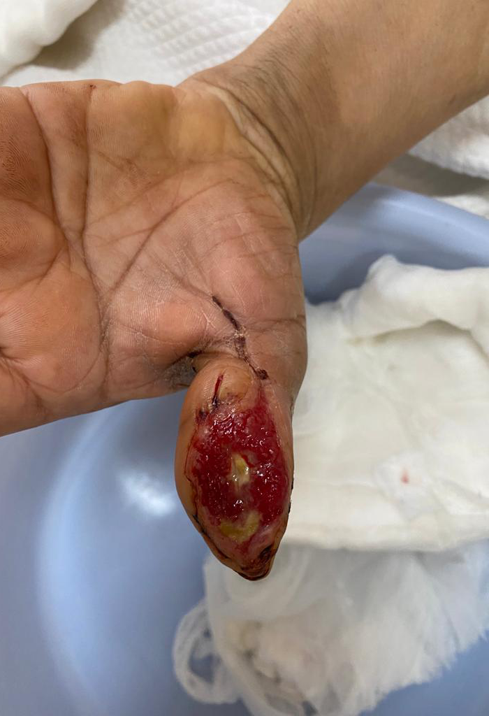
Figure 2. A lesion showing granulation tissue formation after repeated debridement during treatment.
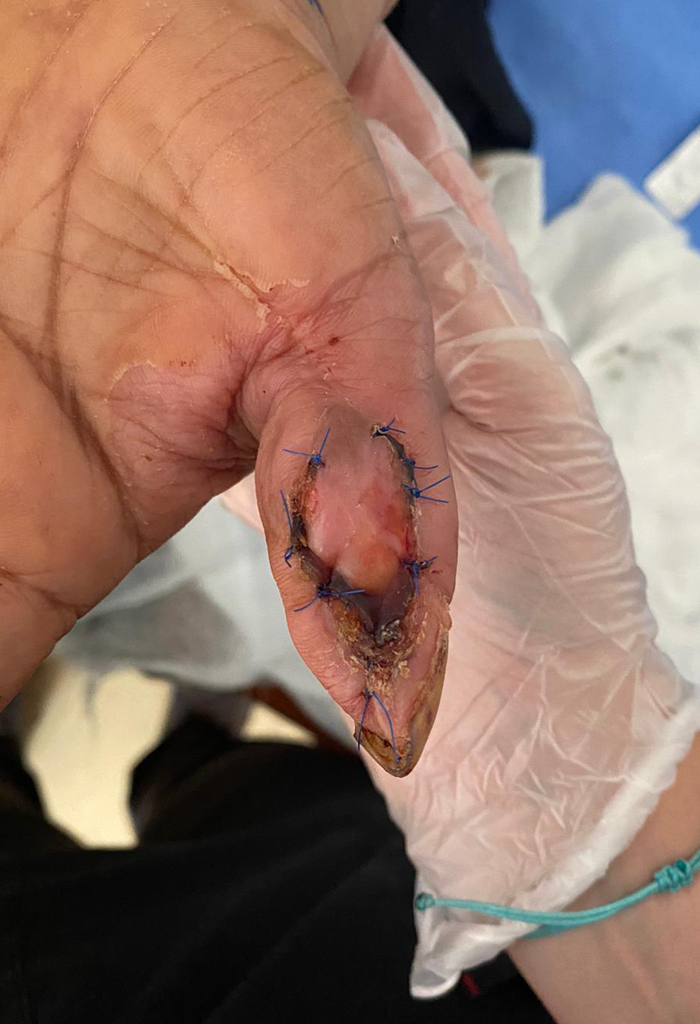
Figure 3. Complete recovery after graft replacement.
Magnetic resonance imaging demonstrated findings consistent with osteomyelitis. The patient required multiple surgical debridements, and a skin graft was applied after adequate granulation tissue formation (Figure 2). The patient has remained free of recurrence for nine months (Figure 3).
Discussion
Diabetic hand infections are rare but serious complications of DM, typically triggered by minor trauma and exacerbated by impaired immune defense. Although more frequently reported in tropical regions, these infections can occur in any geographic setting, particularly in individuals with poorly controlled diabetes and comorbidities (3,4). In the present case, a progressive soft-tissue infection following cactus-thorn trauma was ultimately attributed to C. tropicalis.
Patients with diabetes are predisposed to SSTIs, including fungal infections, through several mechanisms. Hyperglycemia impairs neutrophil function, phagocytosis, and intracellular killing—processes essential for antifungal immunity (5,6). Chronic hyperglycemia also promotes the formation of advanced glycation end products, which compromise the integrity of the skin barrier and promote inflammation (7). Microangiopathy reduces tissue perfusion, while neuropathy leads to delayed recognition of trauma and subsequent delays in seeking care. Together, these factors create a permissive environment for fungal invasion and persistence (1).
Cutaneous invasive fungal infections may arise following environmental contamination of wounds, including those caused by agricultural injuries or thorn punctures. Such infections carry a high risk of morbidity and may require aggressive surgical debridement in addition to systemic antifungal therapy (1,2,5). Although sporotrichosis is classically associated with rose-thorn injury, other fungi have been implicated in similar contexts. For instance, Turkal et al. (8) reported C. parapsilosis infection of the hand following rose-thorn trauma, emphasizing that minor injuries can serve as portals of entry. Villanueva et al. (9) described C. tropicalis cellulitis of the lower limb in a patient with cirrhosis, HIV, and diabetes, underscoring the need for early antifungal therapy in immunocompromised individuals. Tanabe et al. (10) documented Candida albicans flexor tenosynovitis in an immunocompetent patient after minor trauma, demonstrating that Candida spp. can invade the deeper structures of the hand. Wu et al. (11) reported primary cutaneous C. parapsilosis infections in healthy adults and highlighted the diagnostic value of fungal cultures in chronic or atypical skin lesions unresponsive to antimicrobial therapy; all cases responded to itraconazole or fluconazole. Although fungal infections in diabetic wounds are uncommon, they represent a clinically significant cause of morbidity. Early recognition, culture-based diagnosis, timely surgical intervention, and targeted antifungal therapy are critical for achieving optimal outcomes.