Introduction
Crimean-Congo hemorrhagic fever (CCHF) is a zoonotic viral disease caused by the Crimean-Congo hemorrhagic fever virus, a member of the Orthonairovirus genus within the Nairoviridae family, and is associated with high morbidity and mortality (1). Transmission occurs primarily through contact with infected ticks, but exposure to the blood or body fluids of infected animals or patients also represents an important route of infection. In Türkiye, CCHF was first recognized in 2002 and has since remained endemic, particularly in northern Central and Eastern Anatolia and the southern Black Sea region, with a reported mortality rate of approximately 5% (2).
The incubation period generally ranges from two to nine days and depends on the route of exposure, viral load, and host immune status (2). Following incubation, patients typically develop sudden-onset fever, headache, myalgia, arthralgia, nausea, vomiting, and fatigue. In severe cases, hemorrhagic manifestations, coagulopathy, multiorgan failure, and death may occur, with mortality rates reported to reach up to 30% (3).
Crimean-Congo hemorrhagic fever during pregnancy poses a serious threat to both maternal and fetal health. Adverse outcomes such as spontaneous abortion, intrauterine fetal demise, neonatal infection, and maternal death have been reported. Disease severity is a primary determinant of
maternal prognosis, while gestational age at the time of infection primarily influences fetal and neonatal outcomes, including the risk of fetal loss, preterm delivery, and neonatal infection. Vertical transmission may occur via intrauterine or perinatal routes (4).
Case
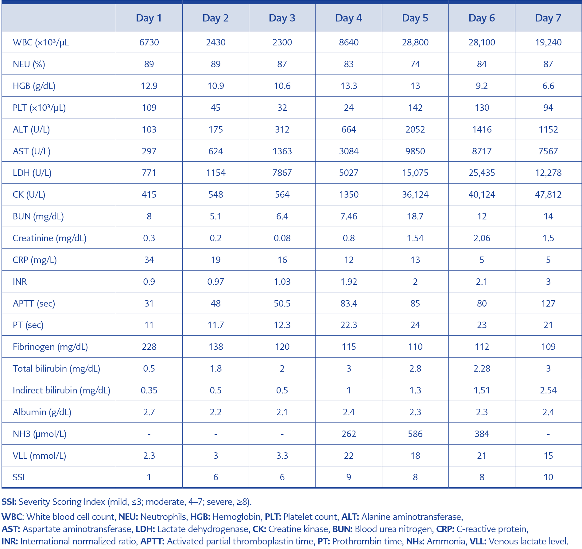
Table 1. Laboratory parameters during hospitalization.
A 30-year-old woman at 23 weeks of gestation with no known chronic medical conditions presented to the emergency department with a five-day history of diffuse body pain, fever exceeding 38°C, and dyspnea. On admission, vital signs were as follows: oxygen saturation 77%, heart rate 108 beats/min, and blood pressure 130/60 mmHg. Initial laboratory findings and Severity Scoring Index (SSI) values, as described by Dokuzoguz et al. (5), are presented in Table 1. Urinalysis revealed 4+ proteinuria. The patient reported regular use of vitamin D and iron supplements and denied using herbal or alternative medications.
She was initially hospitalized in the Obstetrics and Gynecology clinic with preliminary diagnoses of hemolysis, elevated liver enzymes, and low platelet count (HELLP) syndrome and pulmonary thromboembolism. Peripheral blood smear demonstrated giant platelets and marked erythroid poikilocytosis without atypical cells. Pulmonary thromboembolism was excluded by computed tomography pulmonary angiography.
Although there was no history of tick exposure, the patient reported having stayed in a rural area endemic for CCHF approximately 15 days before admission. In addition, she reported recent upper respiratory tract infection-like symptoms in close contacts. Serum samples obtained under appropriate infection-control isolation conditions tested positive for CCHF virus by polymerase chain reaction (PCR). On the third day of hospitalization, the patient was transferred to the Infectious Diseases and Clinical Microbiology Department. She declined ribavirin therapy, and supportive treatment including N-acetylcysteine was initiated.
Chest radiography revealed a right-sided pleural effusion and increased infiltrates in the lower lung zone. Abdominal ultrasonography demonstrated mild gallbladder wall thickening and pericholecystic edema, and empiric meropenem therapy (1 g three times daily) was started. Blood, sputum, and nasotracheal aspirate cultures were obtained prior to meropenem initiation; all cultures showed no growth. Serial fetal ultrasonography showed fetal biometry and heart rate appropriate for gestational age, with no evidence of fetal pathology.
On the fourth day of hospitalization, the patient developed somnolence, hypotension, and tachycardia, accompanied by rapid clinical deterioration. Due to worsening laboratory parameters, platelet transfusion and fresh frozen plasma were administered, along with intravenous methylprednisolone (1 mg/kg). She was transferred to the intensive care unit, where advanced supportive care, including plasmapheresis and hemofiltration, was initiated.
Despite aggressive management, the patient’s condition continued to worsen. On the seventh day of hospitalization, ecchymoses were observed on the trunk and extremities, and laboratory findings were consistent with multiorgan failure and disseminated intravascular coagulation. The patient died, and intrauterine fetal demise was subsequently confirmed.
Discussion
Crimean-Congo hemorrhagic fever is endemic in large regions of Africa, Asia, Eastern Europe, and the Middle East. It is characterized by nonspecific early symptoms followed by hematological abnormalities, including leukopenia, thrombocytopenia, and marked elevations in liver enzymes and markers of muscle injury (4). Physiological immunological adaptations during pregnancy may increase
susceptibility to severe viral infections, although the underlying mechanisms are not fully understood (6).
The clinical course of CCHF in pregnancy is highly variable, ranging from mild disease to fulminant infection resulting in maternal and fetal death. Fatality rates have been reported to be considerably higher in pregnant women and fetuses than in the general population (7). In the present case, the initial clinical and laboratory findings closely resembled HELLP syndrome, which is characterized by hemolysis, elevated liver enzymes, and thrombocytopenia (8). Atypical forms of HELLP syndrome without hypertension or proteinuria may further complicate the differential diagnosis (9).
Several features supported a diagnosis of CCHF rather than HELLP syndrome in this patient, including high-grade fever, diffuse myalgia, acute respiratory compromise, epidemiological exposure in an endemic area, and rapid progression to severe coagulopathy and disseminated intravascular coagulation. In addition, the magnitude and rate of laboratory deterioration exceeded those typically expected in pregnancy-related hypertensive disorders. Notably, gestational age also supported an alternative diagnosis, as HELLP syndrome typically occurs in the third trimester, with second-trimester presentations being exceptionally rare; in this case, the patient presented at 23 weeks of gestation, which further favored an infectious etiology over a hypertensive disorder of pregnancy.
Transplacental transmission of CCHF virus has been documented, and gestational age at infection is a critical determinant of fetal outcome (10,11). In this case, fetal ultrasonographic findings remained normal until maternal multiorgan failure developed, suggesting that fetal demise was closely related to maternal disease severity.
Management of CCHF during pregnancy remains challenging. Ribavirin use is limited due to potential teratogenic effects, and treatment relies primarily on intensive supportive care (12,13). Although plasmapheresis has been associated with clinical improvement in selected severe cases, available evidence is limited to small case series and observational reports predominantly in nonpregnant patients; evidence on its efficacy and optimal timing during pregnancy remains insufficient to draw firm conclusions (14).
Pulmonary involvement is an uncommon but poor prognostic feature of CCHF (15). In this patient, respiratory distress and radiological infiltrates reflected the aggressive multisystemic nature of the disease. Rapid changes in biochemical parameters, including transaminases, lactate, fibrinogen, and platelet counts, were consistent with the fulminant clinical course.
Conclusion
This case demonstrates that CCHF during pregnancy, although rare, may progress rapidly and closely mimic HELLP syndrome, leading to diagnostic challenges and poor outcomes. In endemic regions, pregnant patients presenting with fever, systemic symptoms, and HELLP-like laboratory findings should be promptly evaluated for viral hemorrhagic fevers to ensure early isolation and appropriate management.