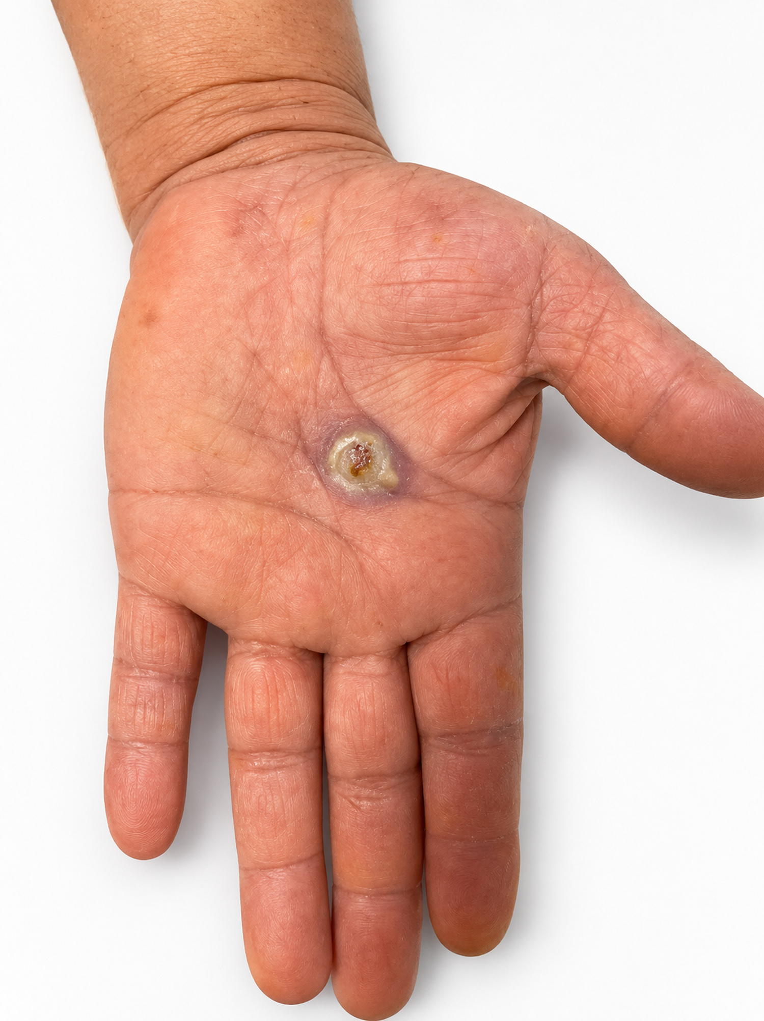
Figure 1. Clinical appearance of the palmar lesion at presentation, showing a solitary, well-circumscribed hyperkeratotic nodule with central ulceration, surrounded by a subtle violaceous halo, with no visible purulent discharge.
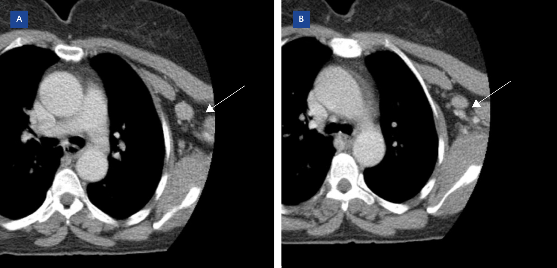
Figure 2. Axial contrast-enhanced CT image showing multiple round and oval left axillary lymph nodes (arrows).
A 55-year-old woman presented with a painful palmar lesion persisting for one year, which had initially appeared as a violaceous papule and gradually evolved into its current form (Figure 1). One week before onset, she removed a thorn-like foreign body from the same site while gardening. Palpation revealed tender ipsilateral epitrochlear and axillary lymph nodes, and computed tomography (CT) confirmed lymphadenopathy at both sites (Figure 2). Histopathology revealed a nonspecific, nongranulomatous dermal infiltrate. Gram staining of the cultured isolate revealed Gram-positive bacilli.
What is the most likely diagnosis?
- Lymphocutaneous sporotrichosis
- Primary cutaneous nocardiosis
- Mycobacterium marinum infection
- Erysipeloid caused by Erysipelothrix rhusiopathiae
All of the listed pathogens should be considered in cases of chronic hand lesions associated with inoculation and regional lymphatic involvement. A history of thorn inoculation and a prolonged clinical course are particularly suggestive of lymphocutaneous sporotrichosis within this spectrum. However, the absence of a characteristic ascending chain of dermal or subcutaneous nodules, together with the painful, nonpurulent nature of the palmar lesion, makes the presentation less typical of lymphocutaneous sporotrichosis (1).
Another consideration associated with soil inoculation is primary cutaneous nocardiosis. However, the absence of suppuration or abscess formation makes this diagnosis less likely. Branching, weakly acid-fast filaments would provide an important microbiological clue.
Mycobacterium marinum is also an important consideration in the differential diagnosis in such cases, as this acid-fast bacillus may appear as a weak or irregular Gram-positive stain on routine Gram staining. However, the absence of exposure to aquatic environments or fish, together with an absence of characteristic ascending nodular lymphangitis, argues against this diagnosis (1,2).
Although not exclusionary, the absence of granulomatous inflammation provides no additional histopathologic support for these alternative diagnoses.
In this patient, the severe pain, nonpurulent palmar lesion, and tender regional lymphadenopathy were clinically consistent with erysipeloid, while histopathology provided no specific support for the principal alternatives. Importantly, culture of a full-thickness biopsy specimen enabled microbiological diagnosis, underscoring the value of deep-tissue sampling. Matrix-assisted laser desorption/ionization time-of-flight mass spectrometry (MALDI-TOF MS) identified the isolate as E. rhusiopathiae, thereby confirming the diagnosis. Accurate identification at the species level is important because the organism may be overlooked as an insignificant coryneform-like Gram-positive bacillus, and it is intrinsically resistant to vancomycin. Amoxicillin, a conventional first-line treatment for erysipeloid, was initiated, and treatment was ongoing at the time of writing (3).