Introduction
Pantoea agglomerans, a Gram-negative, facultatively anaerobic bacterium in the Enterobacteriaceae family, was previously classified as Enterobacter agglomerans or Erwinia herbicola (1). Pantoea agglomerans is not an obligate pathogen for humans. However, two types of infections can occur in humans: 1) skin and joint infections after exposure to plant material, and 2) hospital-acquired infections (2). In this report, we present a case of necrotising fasciitis, initially thought to be a skin and soft tissue infection, in which cultures grew P. agglomerans.
Case
A 52-year-old male patient had screws placed in the lumbar vertebrae following a fall 10 years ago. Subsequently, he developed cauda equina syndrome and was diagnosed with diabetes mellitus about one month prior, which he was managing through diet alone, without any antidiabetic medications. Additionally, he had chronic wounds on the soles of his feet for approximately 10 years.
The patient presented to the emergency department of Ağrı Training and Research Hospital on January 24, 2025, due to the development of a new wound on his left foot over the past week, which had begun to discharge pus. On admission, laboratory tests revealed a white blood cell (WBC) count of 38,000/µL, neutrophils at 36,000/µL, and a C-reactive protein (CRP) level of 360 mg/L. The physical examination showed a body temperature of 36.7°C, a respiratory rate of 22 breaths per minute, and a conscious, oriented, and cooperative state. Widespread rashes were noted on the patient’s left heel and leg. There was no crepitation on palpation. A purulent, foul-smelling discharge was noted from the wound.
The patient was admitted to the infectious diseases service with a preliminary diagnosis of cellulitis and abscess. Given a recent history of cefixime and ciprofloxacin use, empirical broad-spectrum antibiotic therapy with piperacillin/tazobactam and teicoplanin was initiated. A sample of purulent discharge was collected from the new wound on the left foot. A magnetic resonance imaging (MRI) scan was planned due to suspicion of abscess and calcaneal osteomyelitis; however, it could not be performed because the compatibility of the vertebral material was unknown.
Subsequently, superficial ultrasonography (USG) revealed widespread subcutaneous free air in the left lower extremity, raising suspicion for necrotizing fasciitis. The patient underwent bilateral lower extremity computed tomography (CT), which also showed subcutaneous air in the left lower extremity. Based on these findings, teicoplanin and piperacillin/tazobactam were discontinued, and empirical treatment with vancomycin and meropenem was initiated.
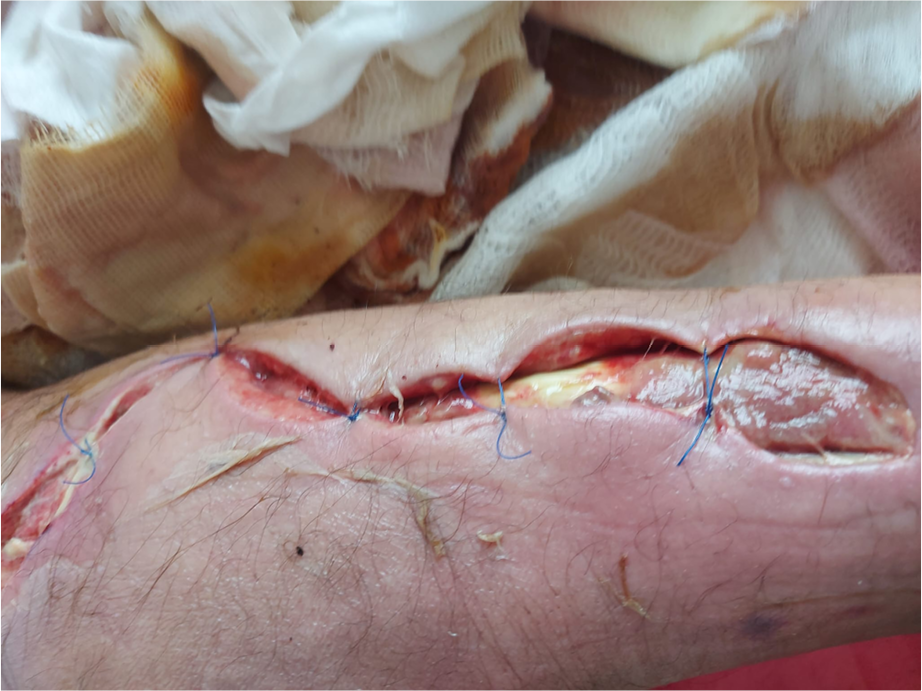
Figure 1. Patient’s leg after debridement.
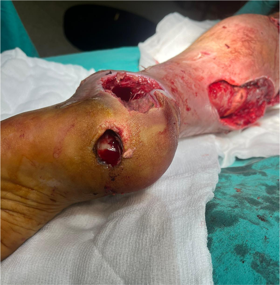
Figure 2. Patient’s foot after debridement.
Plastic and reconstructive surgery was consulted, and on January 28, 2025, the patient underwent extensive debridement with a preliminary diagnosis of necrotizing fasciitis. The diagnosis was confirmed during the operation, with copious purulent discharge drained from the leg. The patient’s leg and ankle are shown postoperatively in Figures 1 and 2, respectively. Samples from the discharge and tissue obtained during surgery were sent for culture. Sheep blood agar and eosin-methylene blue (EMB) agar were used for cultivation. Pantoea agglomerans grew in both preoperative and intraoperative culture samples. Identification and antimicrobial susceptibility testing were performed using the VITEK® 2 Compact system (bioMérieux, Marcy-l’Étoile, France).
Antimicrobial susceptibility testing showed that P. agglomerans was susceptible to amoxicillin/clavulanic acid, cefoxitin, trimethoprim/sulfamethoxazole, amikacin, ceftazidime, ceftriaxone, imipenem, meropenem, cefepime, ertapenem, and ciprofloxacin. It was resistant to piperacillin/tazobactam.
Based on these results, vancomycin and meropenem were discontinued, and ceftriaxone and metronidazole were started. The patient’s wound was dressed, and a second debridement with wound closure was performed by a plastic surgeon, resulting in near-complete wound closure. The patient was given ceftriaxone and metronidazole for 18 days, and he was discharged in full recovery.
Approximately one month later, on April 18, 2025, the patient presented to the plastic surgery outpatient clinic. His leg wound was completely healed, with a small wound remaining on the heel.
Discussion
Necrotizing fasciitis is a rare, rapidly progressive soft tissue infection that can lead to sepsis and death (3,4). It is classified into two types: polymicrobial (type 1) and monomicrobial (type 2). Type 1 infection is more common and is usually caused by Gram-positive microorganisms (such as Staphylococcus aureus, Streptococcus pyogenes, and enterococci), Gram-negative aerobes (such as Escherichia coli and Pseudomonas spp.), and anaerobic microorganisms (Bacteroides spp. and Clostridium spp.) are usually the causative agents. Most type 2 infections are caused by Gram-positive microorganisms, particularly S. aureus and streptococci (5).
Pantoea agglomerans is not typically an obligate infectious agent in humans. It can cause opportunistic infections in two main situations: 1) wound infections associated with exposure to plant material, and 2) hospital-acquired infections. Wound infections with P. agglomerans may occur following skin puncture or laceration by plant thorns, wooden splinters, or other plant material, commonly during agricultural work, gardening, or while children are playing (2).
In our case, P. agglomerans was identified as the causative agent through tissue cultures. Although soft tissue infections and bacteremia due to P. agglomerans have been reported (6,7), to our knowledge, this is the first case of necrotizing fasciitis caused by
P. agglomerans reported from Türkiye.
Conclusion
Necrotizing fasciitis is a rare but life-threatening infection, and obtaining appropriate culture specimens is crucial for accurate diagnosis and effective treatment.