Introduction
Historically, Mycoplasma hominis was the first mycoplasma species isolated from humans, first identified in 1937 from a Bartholin’s gland abscess. It is a small, coccoid-shaped (approximately 0.5 μm), Gram-negative bacterium that grows slowly in culture (1). Due to the lack of a cell wall, M. hominis is resistant to beta-lactam antibiotics (2). Treatment typically includes tetracyclines, fluoroquinolones, or clindamycin (1).
Mycoplasma hominis is commonly found as a commensal organism in the urogenital tract, with a reported prevalence of 21–53% in individuals, depending on socioeconomic status and sexual activity (1,3). More recent studies have reported prevalence rates ranging from 0%–17% in the female genital tract and 4%–7% in the male genital tract (3,4). Kim et al. (5) found M. hominis colonization in 15% of asymptomatic, sexually active women and 9% of men. Additionally, M. hominis has been detected in respiratory tract secretions, with a prevalence of 2.2% in men and 0% in women (4).
Although M. hominis rarely causes clinical infections in immunocompetent individuals, it can lead to various urogenital infections, including urinary tract infections, bacterial vaginosis, pelvic inflammatory disease, and pregnancy-complications. In immunocompromised patients or following surgical procedures such as organ transplantation or prosthesis implantation, it can also cause extragenital infections, including central nervous system infections, septic arthritis, endocarditis, and mediastinitis (1,3). Reports indicate that the incidence of pharyngitis due to M. hominis ranges from 0.5 to 16.6% (3), and M. hominis has been isolated from pharyngeal secretions of 14.3% of asymptomatic women with a history of oral sex (6).
To date, only one case of peritonsillar abscess due to co-infection of M. hominis and Fusobacterium necrophorum has been reported. That case involved a 19-year-old healthy female patient following oral sex, raising questions about the true etiologic role of M. hominis (3,7).
Here, we present the case of a healthy 27-year-old woman who developed a left peritonsillar abscess following sexual intercourse. To our knowledge, the present case represents the first documented instance of a peritonsillar abscess solely caused by M. hominis following sexual intercourse.
Case
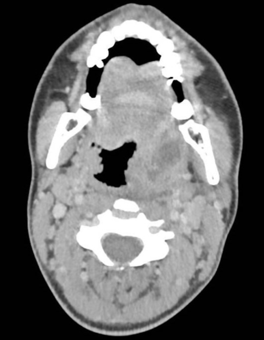
Figure 1. Cervical computed tomography (CT) scan showing a left peritonsillar abscess.
A 27-year-old previously healthy female was referred to our tertiary care center from a local clinic with a diagnosis of a left peritonsillar abscess confirmed by computed tomography (CT) (Figure 1). She reported a two-week history of odynophagia, which had started two weeks after a sexual encounter. Her symptoms have worsened over the last three days, with the onset of a dysphonia and trismus.
Upon presentation, the patient appeared to be in stable condition, and her vital signs were within normal limits. Physical examination revealed bilateral cervical adenopathy, mild trismus (two fingerbreadths), and swelling of the left soft palate with slight deviation of the left tonsil towards the medial pharynx. Laboratory tests showed leukocytosis (14.5 × 10⁹/L) and an elevated C-reactive protein (CRP) level of 106 mg/L. Empirical intravenous antibiotic therapy with co-amoxicillin (2.2 g three times daily) had already been initiated prior to hospital transfer.
A quinsy tonsillectomy was performed on the left side without complication. The pus from the abscess cavity was sent for microbiological analysis. The antibiotic therapy was continued, and the patient showed rapid improvement, leading to hospital discharge 48 hours after surgery.
Anaerobic culture of the aspirated pus yielded M. hominis at a concentration of 103 CFU/mL. Based on the clinical timeline, the infection was presumed to have been sexually acquired. The pathology report of the left tonsil indicated signs of tonsillitis, including lymphoid hyperplasia and acute peri-tonsillitis.
At a follow-up visit, we conducted a sexually transmitted infection (STI) check-up, including tests for HIV, hepatitis B and C, syphilis, and oropharyngeal swabs for Chlamydia trachomatis and Neisseria gonorrhoeae; all of which returned negative results. We also recommended a gynecology examination. The swab for M. hominis remained positive, although the patient was asymptomatic with normal oropharyngeal findings. Following consultation with an infectious disease specialist, no further antibiotic treatment was recommended.
Discussion
This case highlights a rare instance of a peritonsillar abscess caused solely by M. hominis, occurring after sexual intercourse.
Three prior case reports have documented deep neck infections caused by M. hominis in previously healthy individuals. The first, published in 2008, described a parapharyngeal abscess following acute Epstein-Barr virus infection in a 20-year-old male (8). The second, in 2016, involved a case of mediastinitis secondary to a tonsillar abscess in a 59-year-old male; M. hominis was isolated despite the initial causative organisms being undocumented (2). The third case, reported in 2023, described a 19-year-old woman who developed a peritonsillar abscess after sexual intercourse, with a co-infection of M. hominis and Fusobacterium necrophorum (3). Notably, that report was the first to suggest a potential link between sexual activity and M. hominis-related peritonsillar abscess (3).
In contrast, our case is unique in that M. hominis was the only pathogen identified from the peritonsillar abscess, strongly implicating it as the primary causative agent. This suggests that M. hominis can independently cause a peritonsillar abscess following sexual intercourse.
Conclusion
Larger studies are needed to determine the true prevalence of M. hominis in deep neck infections. It is important to include a sexual history in the evaluation of all patients presenting with a peritonsillar abscess.